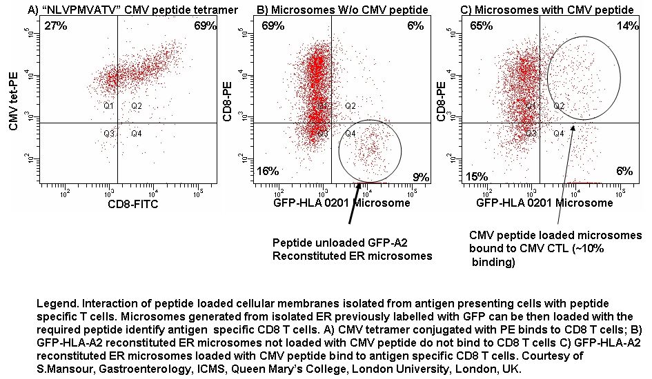
Isolation of GFP-HLA-A2 labelled Endoplasmic Reticulum (ER) can be sonicated into microsomes and identified by flow cytometry and loaded with CMV-peptide to identify antigen specific CD8-PE T-cells, see figure.
|
|
|||||||||||||||
| |
|||||||||||||||
 |
|||||||||||||||
| |
|||||||||||||||
|
|||||||||||||||
|
|||||||||||||||
| by Gary Warnes. © Queen Mary, University of London 2007 | |||||||||||||||
| Institute of Cell and Molecular Science, Barts and The London, Queen Mary's School of Medicine and Dentistry, Queen Mary, University of London, 4 Newark Street, London E1 2AA, Tel: +44 (0)20 7377 7000, Fax: +44 (0)20 7247 3428 | |||||||||||||||