Tinctorial Stains
Biological tissue has little inherent contrast in either the light or electron microscopy. Staining is employed to give both contrasts to the tissue as well as highlighted particular features of interest.
Tinctorial staining (or more commonly known as special staining) has been used to selectively stain cells and cellular components. A variety of compounds used to colour tissue sections include, Safranin, Oil Red O, Congo Red, Fast Green FCF, Silver Salts and numerous natural and artificial dyes that were usually originated from the development of dyes in the textile industry.
Histochemistry refers to the science of using chemical reactions between laboratory chemicals and components within tissue. Below is a list of the most commonly used stains in a laboratory’s repertoire. If you do not see what you require please contact a member of staff as many of the stains can be adapted and there are more available.
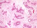
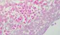
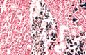
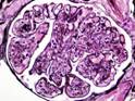
Haematoxylin and Eosin Stain (H&E) Gills haematoxylin and Eosin is the standard method I the lab but other stains; like Ehirlichs and Weigert’s haematoxylin for nuclei are also available. Nuclei stain blue, other elements shades of pink/red. |
|
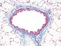
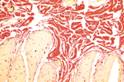
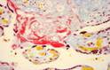
Connective Tissue Stains |
|
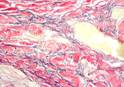
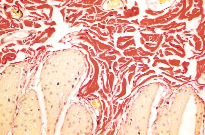
Masson Trichrome |
|
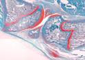
Van Gieson |
|
MSB |
|
Elastic Van Gieson (EVG) |
|
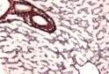
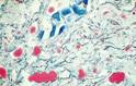
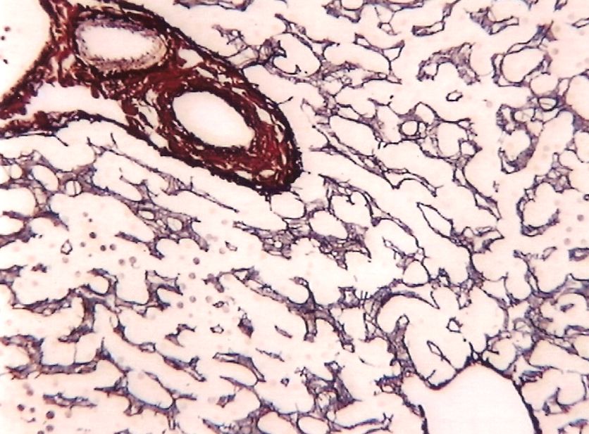
Gordon & Sweet’s Stain for Reticulin Fibres |
|
Safranin O |
|
Nucleic Acids |
|
Feulgen Stain |
|
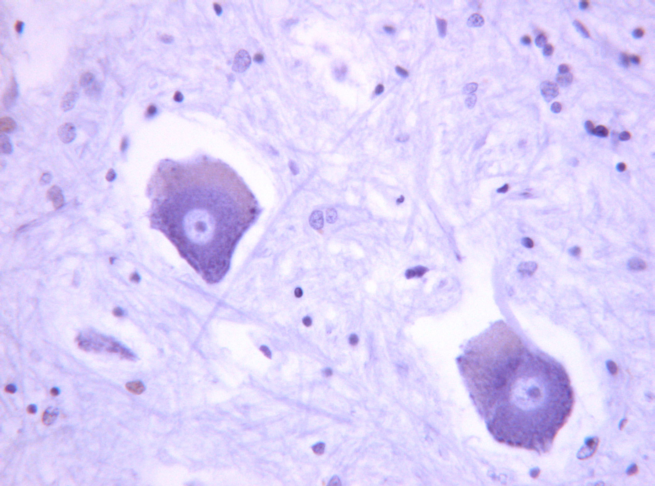
Methyl Green – Pyronin |
|
Amyloid |
|
Congo Red |
|
Glycogen, Carbohydrates and Mucins |
|
Periodic Acid Schiff (PAS) |
|
Alcian Blue for Acid Mucins |
|
Lipids |
|
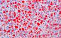
Oil Red O |
|
Nile Blue |
|
Pigments & Minerals |
|
Perls’ Prussian Blue |
|
Fouchet |
|
Masson Fontana |
|
Von Kossa |
|
Rhodanine |
|
Grimelius |
|
Hexamine Silver |
|
Luxol Fast Blue Cresyl Fast Violet |
|
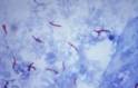
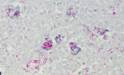
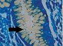
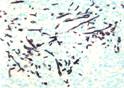
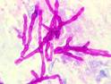
Micro Organisms and Fungi |
|
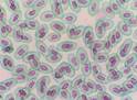
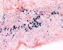
Gram Stain |
|
Ziehl-Nelson |
|
Wade-Fite |
|
Alcian Yellow Toluene Blue |
|
Grocott |
|
PAS |
|