Electron Microscopy
Introduction
To complement the excellent service in materials sciences provided by NanoVision
( http://www.nanovision.qmul.ac.uk/ ) Centre which is headed by Dr. Bushby (a.j.bushby@qmul.ac.uk), the Blizard Institute Core Pathology has teamed up with the EM department of the Barts and the London Hospital to provide an EM biological sample preparation service.
The EM service at the Cellular Pathology department of the Royal London Hospital is headed by Mr. Graham McPhail who has years of EM experience and is regarded as an expert in his field.
The Service
The department has the ability to process all types of samples for both Transmission EM and Scanning EM. The samples range from;
- Agarose gel samples containing cell suspensions
- Cover slips with a monolayer of cells
- Tissue samples both human and animal
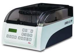
Where suitable the processing is carried out on our Leica EM tissue processor. The processor is designed for EM resin processing, when this is not suitable then we will carry out the processing by hand.
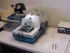
We also cut both ultra thin and semi thin sections and stain them for EM imaging.
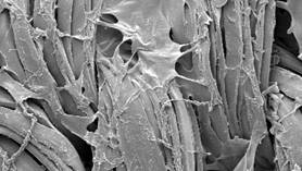
We will also image the sections for you on the EM microscope based at NanoVision and provide digital images on a CD. We can either do this for you, you can attend the session and choose the areas you would like imagin. Teaching of the use of the EM microscope can also be given.
Contact
For further information please contact either Mr. Evagora or Mr. McPhail.
Christopher Evagora – Laboratory Manager of Core Pathology
Telephone: 020 324 60155
E-mail: c.a.evagora@qmul.ac.uk
Graham McPhail – head of EM Service at Barts and the London Hospital
E-mail: Graham.McPhail@bartsandthelondon.nhs.uk
Cost
This a fee-for-service set up and the prices are outlined below.
Service |
Cost (per specimen) |
Fixation, processing and embedding in Araldite |
£ 10.00 |
Block trimming and cutting of semi-thin sections from initial level of block. Staining of sections with toluidine blue |
£ 7 .00 |
Cutting and staining semi-thin sections from additional levels (per block, per level) |
£ 4.50 |
Cutting ultra thin sections and mounting them on 2-4 grids (per block, per level) |
£ 12.50 |
Staining of ultrathin sections with uranyl acetate and lead citrate (up to 4 grids) |
£ 7.00 |
Examination of specimen in electron microscope (per hour) |
£ 25.00 |
Training in electron microscopy techniques and / or use of electron microscope (per hour) |
£ 25.00 |
Digital electron micrographs on CD-ROM (per disk) |
£ 1.00 |
Preparation of SEM samples |
£ 10.00 |
Please not an additional charge may be made by Nanovision for the use of the electron microscope.