
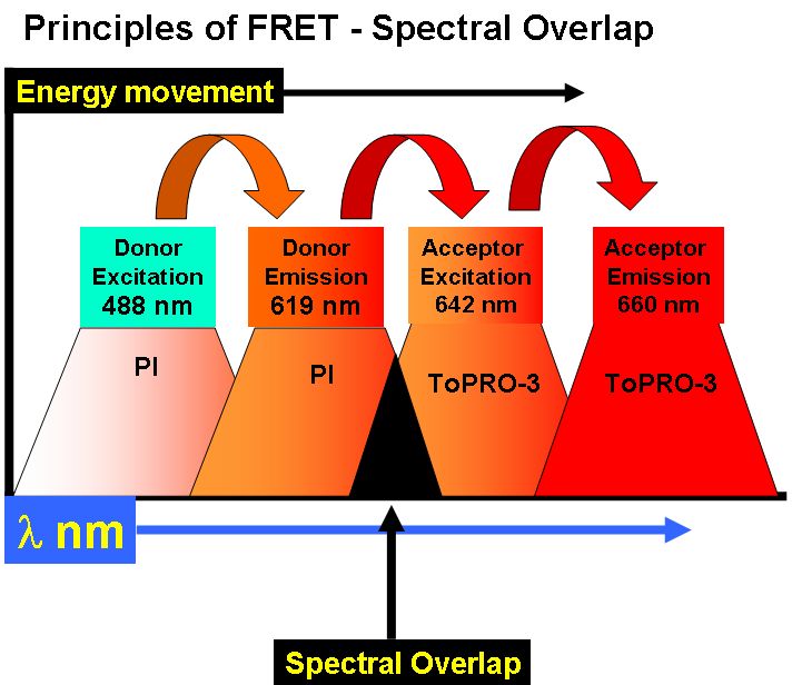
Fluorescence Resonance Energy Transfer or FRET can be used to show that molecules are within 10 nm of each other by nature of the transfer of energy via a long range dipole-dipole coupling mechanism. A fluorescent donor molecule is excited by a light source, the energy is nonradiatively transferred to an acceptor molecule, see figure Principles of FRET.
The efficiency of this energy transfer depends upon the distance between acceptor and donor, the degree of spectral overlap between the emission spectra of the donor and absorption of spectra of the acceptor and the relative parallel orientation of donor and acceptor dipoles. The distance at which the energy transfer is 50% efficient is called the Forster Radius (Ro). The magnitude of the Forster Radius for each fluorophore is also dependent upon the relative quantum yield of the fluorophore.
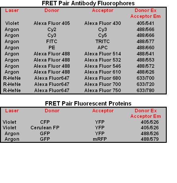
FRET Pairs
Thus the choice of fluorophores for a FRET experiment, or FRET pairs of donor and acceptor molecules is important given the range of spectral properties of fluorophores. Originally FITC & TRITC was used as FRET pairs. More recently the Cy dyes have been used as FRET pairs e.g. Cy2-Cy3 or Cy3-Cy5. The Alexa Fluor Dyes also act as FRET pairs e.g. Alexa Fluor 488 and Alexa Fluor 546. Fluorescent Proteins such as CFP-YFP, GFP-YFP and GFP-mRFP also act as FRET pairs, see table for more detailed information.
FRET can used to measure BrdU incorporation by the use of DNA dyes PI and ToPro-3.
FRET based gene reporter systems
A gene reporter system developed by Invitrogen uses FRET-based fluorescent substrates, CCF2 and CCF4.
Fluorescent Protein based FRET
Fluorescent proteins (FPs) can also be used to detect protein interactions or biological processes such as phosphorylation. The most commonly used FRET pairs of FPs are CFP-YFP, but GFP-YFP and GFP with DsRED, mRFP or RFP can also be used in flow cytometric detection of FRET. The development of gene transfection for single proteins expressing CFP and YFP at terminal ends of the protein allow FRET to occur when the protein undergoes a conformational change when acted upon by a specific biological process e.g. phosphorylation. CFP-YFP FRET can be detected by the Core Facilities LSR II analyser and Aria cell sorter enabling the sorting of FRET expressing cells.