
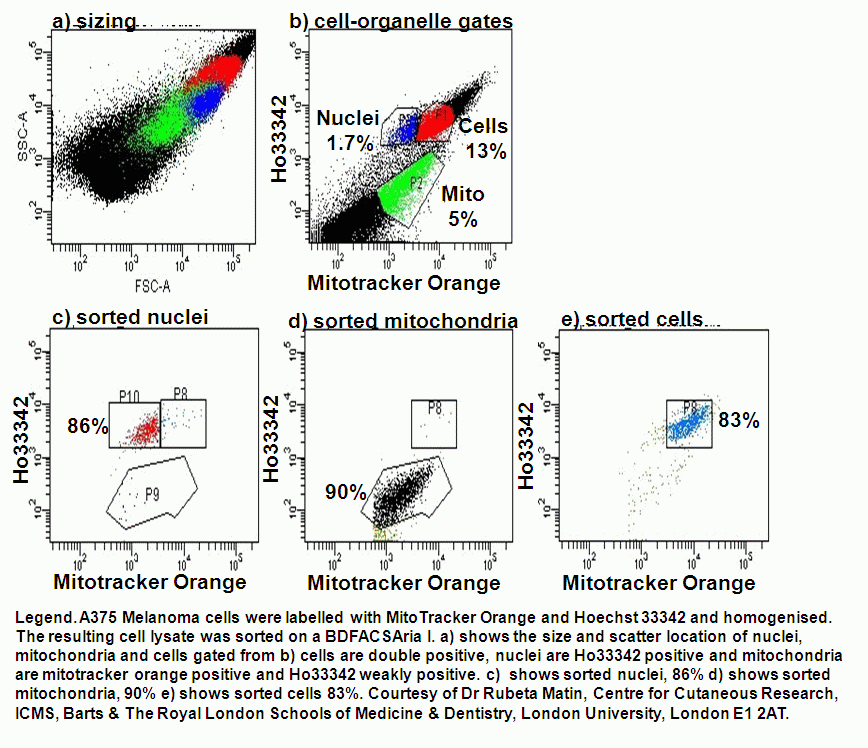
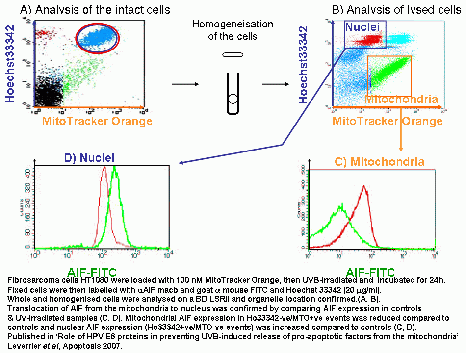
The translocation of proteins involved in apoptosis can be semi-quantitated by flow cytometry. Fluorescently labelling the organelles and the protein under investigation allows the protein to be measured by flow cytometry after lysing the cell population.
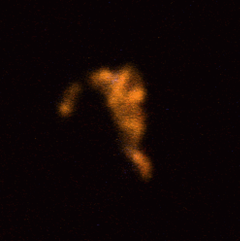
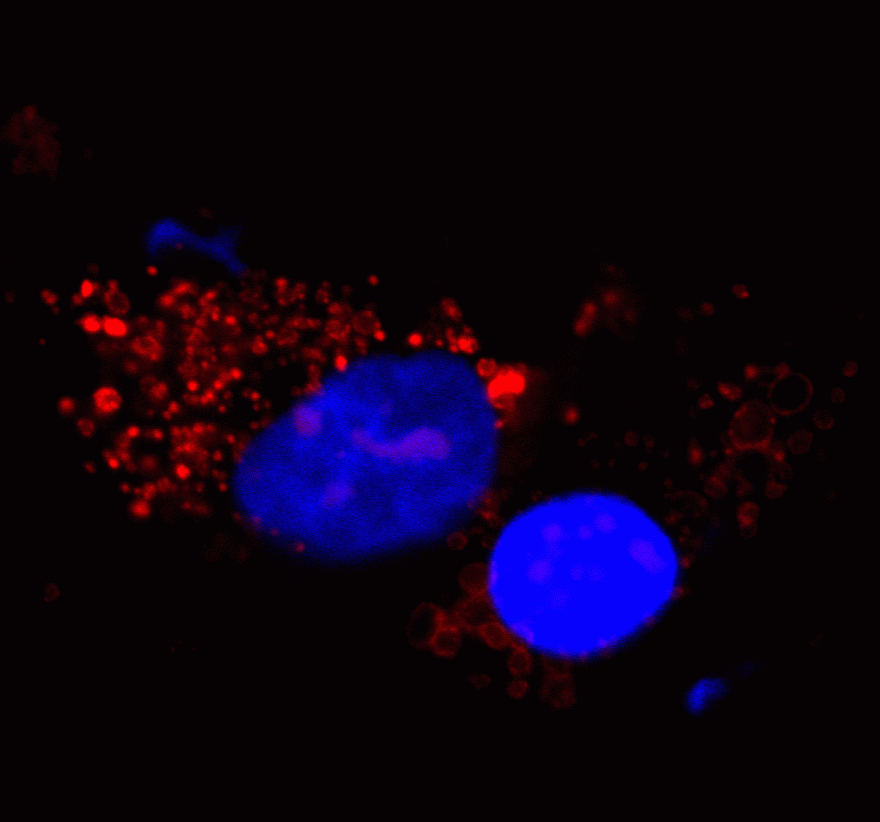
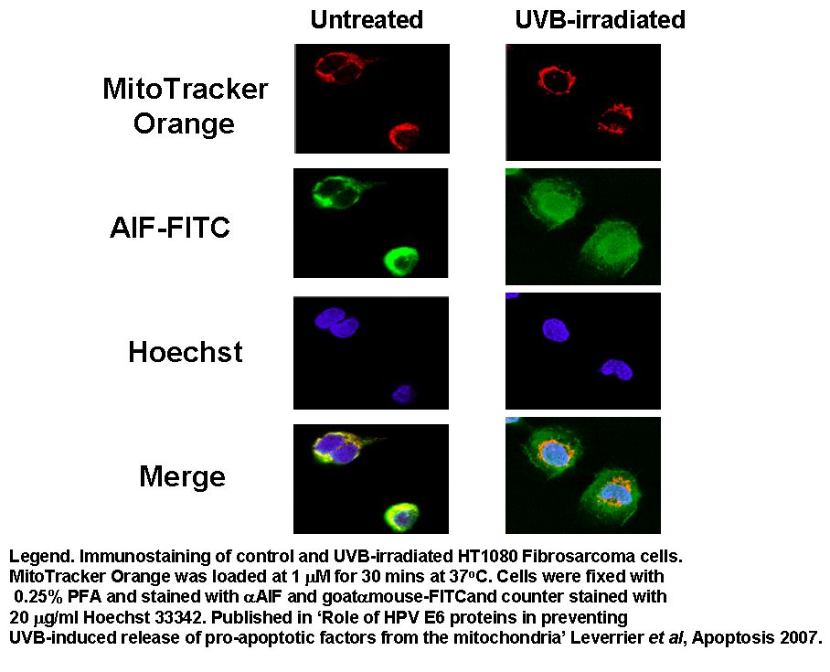
Apoptosis Inducing Factor (AIF) translocates from the mitochondria to the nucleus in cells undergoing apoptosis where AIF fragments DNA. This translocation can be measured by fluorescently labelling mitochondria with Mitotracker Orange, this probe is not released upon cell death or fixing of the cells. Cells were then UVB-irradiated and incubated for 24 h.
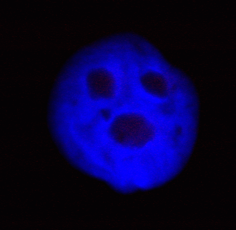
Cells were then fixed and permeabilised and labelled with anti-AIF-FITC and Hoechst 33342. Whole cells are analysed and confocal laser scanning micrographs taken to show the intracellular location of the fluorescent probes. After homogenization, cells were re-analysed on a BD LSRI with 1 million events collected.
Cells and organelles were also sorted according to Ho33342 and MitoTracker Orange staining. Nuclei are Ho33342+ve MitoTracker Orange-ve, Mitochondria are Mitotracker Orange+ve-Ho33342-weak+ve and cells double positive. Purities were checked after sorting and all showed over 80% purity. Confocal imaging of sorted fractions showed that the cells were intact, nuclei were free of mitochondria and cytoplasm and mitochondria showed no nuclear material to be present. Sorting the organelles studied confirms that proteins detected in the fractions by flow cytometric analysis are organelle specific, see figure.
Publications.
RN Matin, A Chikte, SLP Chong, D Mesher, M Graf, P Sanza, V Senatore, M Scatolini, F Moretti, IM Leigh, CM Proby, A Costanzo, G Chiorino, R Cerio, CA Harwood, D Bergamaschi. p63 is an alternative p53 repressor in melanoma that confers chemoresistance and a poor prognosis. J Exp Med Feb, 210 (3), 581-603, 2013.
S Leverrier, D Bergamaschi, L Ghali, A Ola, G Warnes, B Akgul, K Blight, R Garcia-Escudero, A Penna, A Eddaoudi, A Storey. Role of HPV E6 proteins in preventing UVB-induced release of pro-apoptotic factors from the mitochondria. Apoptosis 12 (3), 549-60, 2007.