

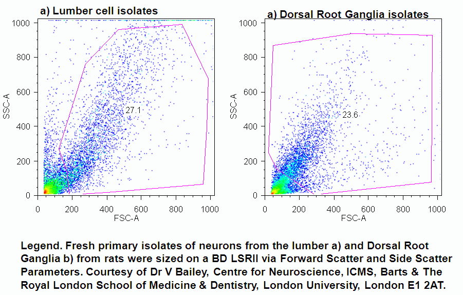
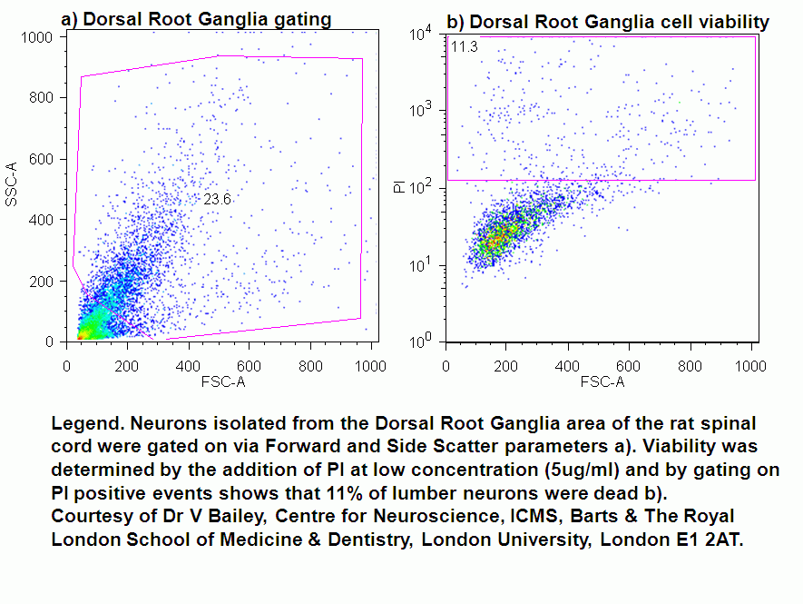
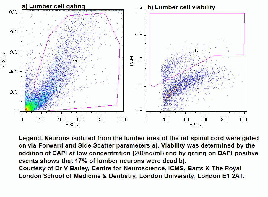
The rat is used in neuroscience as an animal model in the study of spinal cord repair. Researchers extract neuronal cells from the lumbar and dorsal root areas of the spinal cord. Flow cytometry can be used to determine neuronal cell viability after extraction from the animal.
Flow cytometric analysis of primary neurons
Adjustment of Forward scatter and side scatter voltages shows the presence of the large neurons from the Lumber and Dorsal Root areas of the spinal cord. There is a large amount of debris from these primary isolated cells with only 20,000 neurons present from a single spinal cord extract.
Viability
After gating on the primary neurons via forward scatter and side scatter viability of these fresh primary neural isolates can be determined by the use of propidium iodide (PI) at 5ug/ml and DAPI at 200 ng/ml.