
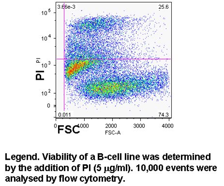
Cell viability can easily be determined in flow cytometry by adding one of DNA binding dyes at relatively low concentration to a population of cells. A common approach is to use propidium iodide (PI) and 5 mg/ml; but there is a wide selection of dyes that can be used, which include 7-AAD, DAPI, DRAQ7 (Biostatus), SYTOX ADDVanced and To-Pro-3 all of these have to be used with caution as live cells will eventually take up these dyes if left to long, see figure and protocols.
Determination of cell viability after fixing
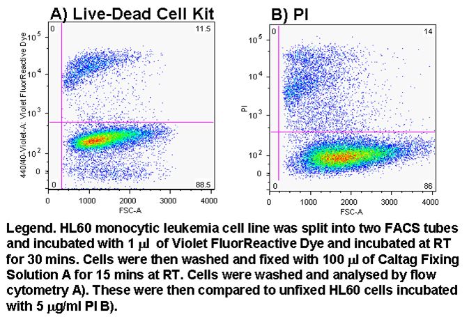
The determination of cell viability when cells are infected with a pathogenic organism can be problematic given the requirement to fix the sample before flow cytometric analysis. However using the new Invitrogen reagent Violet Fluor-Reactive Dye allows the investigator to determine cell viability by flow cytometry by pre-incubation of cells with dye before fixing, see figure and protocol.
The NEW Biolegend Zombie dyes are a cheaper alternative to Invitrogen's fixable dyes. There are 3 colour Zombies available from Near Infra Red (NIR) which is sub-optimally excitable by a red laser (633nm, Ex max 716nm) and emits at 746nm or in the APC-Cy7 channel, see figure. The compensation in the other red channels such as APC & Alexa Fluor-700 is only possible with over 100% compensation and is therefore not recommended. The other two Zombies Dyes, Aqua & Yellow are both excited by UV and Violet lasers with maximal excitation at 382 & 396nm respectively. The emission of Zombie Aqua & Yellow are 510 and 572nm, so FITC and PE filters were placed on the UV or Violet Trigon (BD instruments). The bleed through of Zombie Yellow into the 510 is higher (making it unusable) than that of the Pacific Blue Channel (440/40nm) making it possible to use these two dyes even without a UV laser. UV laser detection of Aqua or Yellow Zombies allows the removal of their emissions on violet laser detector arrays making it possible to combine Zombie fixable dyes with BV421 (Pac Blue), BV501 & BV570.
Millipore Muse cell counting and viability
Cell counts per volume and cell viability can be determined on the mini flow cytometer the Millipore Muse. Cells to be counted are mixed with the cell viability dyes supplied in the Cell Count and Viability Kit (Cat No MCH100103). Small debris is then gated out via a cell size parameter. Dead cells are double positive whilst live cells are single positive as shown on the cell nucleated parameter. Dilution factors can be inputted into the software to show the cell concentration in the original sample, see figure.
Early and late stages of cell death
A cell becomes necrotic when the plasma membrane integrity is compromised sufficiently to let viability dyes such as propidium iodide, DAPI or DRAQ7 into the cell. Late apoptosis or necrosis can be identified by SubG1 DNA analysis. Early necrosis can now be identified by the use of plasma membrane probe, Bis-oxonol. Dead or necrotic cells display a differential signal of Bis-oxonol. DNA analysis of high and low intensity Bis-oxonol events show that low intensity events have a greater level of DNA in the SubG1 area compared to the high intensity events, see figure. The side scatter of high and low intensity bis-oxonol events are also larger when the intensity of bis-oxonol is high and vice a versa. Thus dead cells can now be identified as early or late necrosis by a combination of a viability dye, Bis-oxonol,The side scatter of high and low intensity bis-oxonol events are also larger when the intensity of bis-oxonol is high and vice a versa. Thus dead cells can now be identified as early or late necrosis by a combination of a viability dye, Bis-oxonol, quantification of DNA and side scatter, see figure.
