
Necrosis is the process occurring in cells that lack plasma membrane integrity. Thus necrosis can be derived from apoptosis, oncosis or accidental cell death, or autophagy or self phagocytosis.
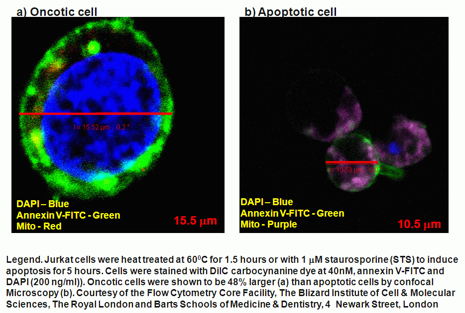
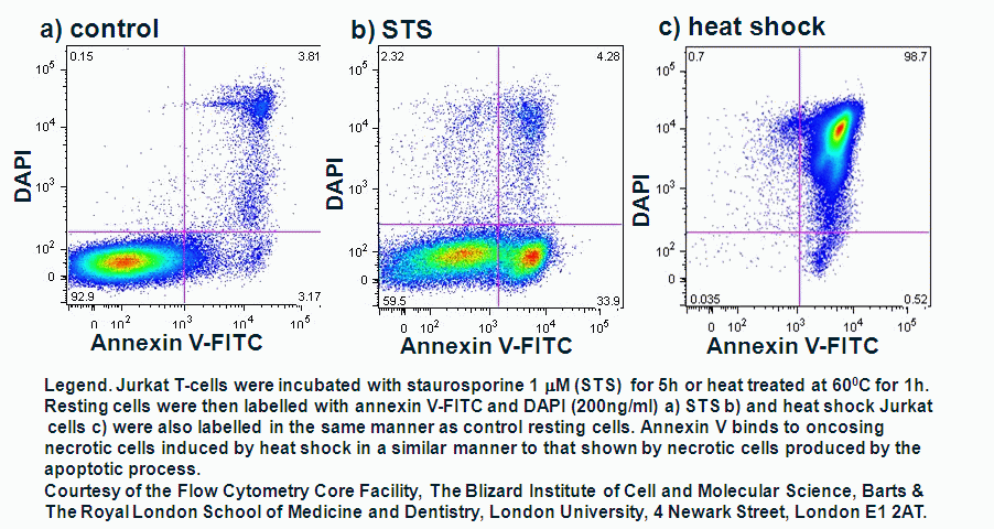
Oncosis is the induction of necrosis by a nonphysiological event resulting in cell swelling as opposed apoptosis where there id cell shrinkage. Oncosis is induced by heat shock (56C or 42C), sodium azide (1% solution), Triton X-100 (0.01% solution) or drugs used to induce apoptosis at a higher concentration. Necrotic cells can be derived from the induction of apoptosis, oncosis or autophagy and refers to the necrobiological events after loss of plasma membrane integrity. During oncosis the cell swells as does cell organelles such as mitochondria but the plasma membrane remains intact. Although it has been reported that phospholipid asymmetry is lost and the externalized phosphatidylserine can bind annexin V this is not part of the oncotic process, see figure. This is followed by the rupture of the plasma membrane during oncosis or the necrotic stage of oncosis leads to the release of proteolytic enzymes causing damage to the surrounding tissues. During apoptosis the cell shrinks in size, mitochondrial structure changes with loss of transmembrane potential, loss of asymmetry of phospholipid's and condensation of chromatin, See section on Apoptosis. This is followed by membrane blebbing and formation of apoptotic bodies and the cell is then necrotic but derived from an apoptotic process not oncosis, see figure for comparison of oncotic and apoptotic cells.
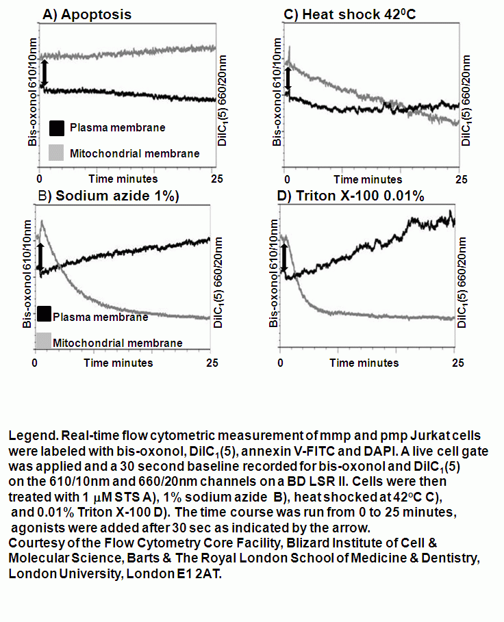
Real-time membrane potential changes for the kinetic analysis of oncosis
Apoptosis is an ATP dependent process whilst oncosis is not, this fact can be used to differentiate between the two processes in a real-time manner by flow cytometric analysis of mitochondrial membrane potential by use of carbocyanine dye DiIC1(5). The different effects induced by the oncotic and apoptotic processes upon cell membranes of the cell this is especially true of the plasma membrane. The potential of the plasma membrane can be measured in a real-time manner by use of the Oxonol dye, DiCAB4 or bis-oxonol, see figure.
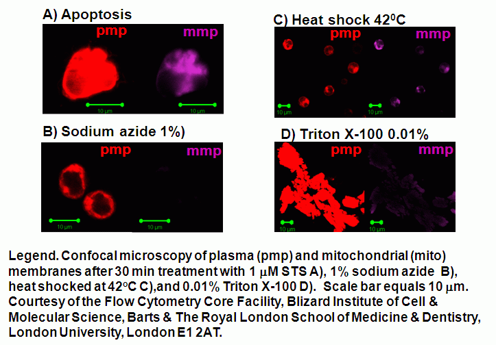
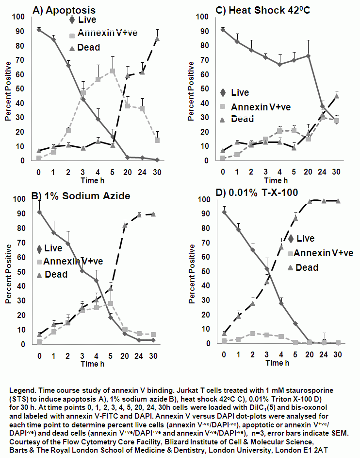
Oncotic agents, such as heat shock, sodium azide and Triton X-100 induce a rapid detectable reduction in mitochondrial function and plasma membrane depolarization; as shown by a rapid fall and increase in DiIC1(5) and bis-oxonol intensity respectively. Whereas induction of apoptosis results only in small insignificant changes in membrane potentials. These changes in membrane potentials during oncosis can be observed by confocal microscopy, see figure.
Plasma membrane potential changes during oncosis
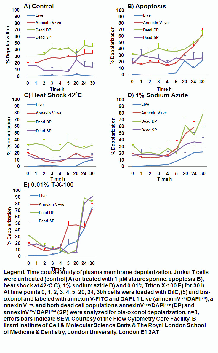
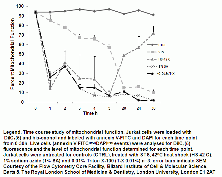
The rapid changes in plasma membrane potential observed during the initial stages of oncosis can also be measured in a time dependent manner over a 30 hour period in a similar manner to that of annexin V binding. A higher proportion of cells undergoing oncosis show depolarization of the plasma membrane, especially cells that are dead and bind annexin V; as does annexin V binding cells, dead cells that do not bind annexin V and even live cells although these do maintain membrane integrity they have little mitochondrial function, see figure. See also the section Viability on the role of plasma membrane depolarization in phases of cell death.
Detection of Early and Late Necrosis
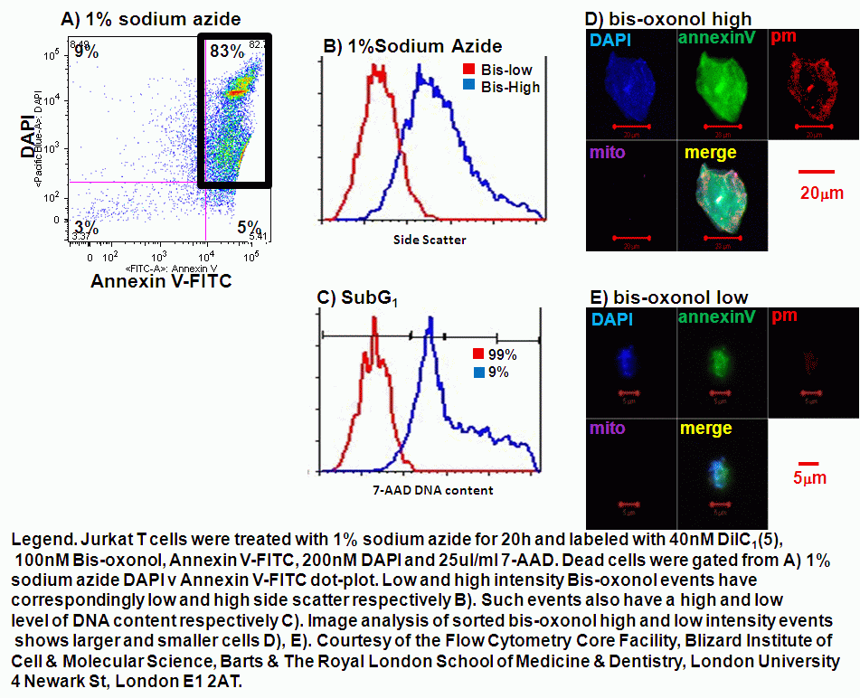
A cell becomes necrotic when the plasma membrane integrity is compromised sufficiently to let viability dyes such as propidium iodide, DAPI or DRAQ7 into the cell. Late apoptosis or necrosis can be identified by SubG1 DNA analysis. Early necrosis can now be identified by the use of plasma membrane probe, Bis-oxonol. Dead or necrotic cells display a differential signal of Bis-oxonol. DNA analysis of high and low intensity Bis-oxonol events show that low intensity events have a greater level of DNA in the SubG1 area compared to the high intensity events, see figure. The side scatter of high and low intensity bis-oxonol events are also larger when the intensity of bis-oxonol is high and vice a versa. Thus dead cells can now be identified as early or late necrosis by a combination of a viability dye, Bis-oxonol, quantification of DNA and side scatter, see figure.
- Model of plasma membrane
- Comparison of necrotic and apoptotic cell size
- Confocal microscopy of oncosis
- Annexin V protocol
- Mitochondrial membrane dye protocol
- Plasma membrane dye protocol