
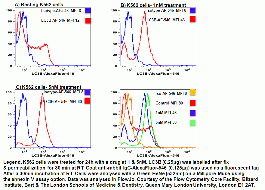
The micro-tubule associated LC3-II(B) is the main biological marker for autophagy. LC3-I is located in the cytoplasm and is moved to the phagosome during autophagosome formation where it is lipidated by the phospholipid, phospha-tidylethaloamine (PE) to form LC3-II(B). Rabbit polycolonal antibodies are often used in the detection of LC3B as a marker of autophagy for image and more recently flow cytomtric analysis. This antibody is non-species specific reflecting the highly conserved nature of this protein this can be fluorescently tagged by indirect immunostaining. LC3B was tagged with AlexFluor-546 which is optiamlly excited by the Muse Green HeNe laser and can be manually analyzed using the Muse Annexin V protocol Yellow parameter, see figure.
Alternatively there is a non-LC3B specific kit from EMD Millipore’s Muse™ Autophagy LC3-antibody based kit provides a quantitative solution for the study of autophagy. This kit contains two key detection reagents to help facilitate the monitoring of lipidated LC3-II in a given cell system:
1) The use of selective permeabilization solution discriminates between cytosolic LC3 from autophagic LC3 by extracting the soluble cytosolic proteins, while protecting LC3 which has been sequestered into the autophagosome;
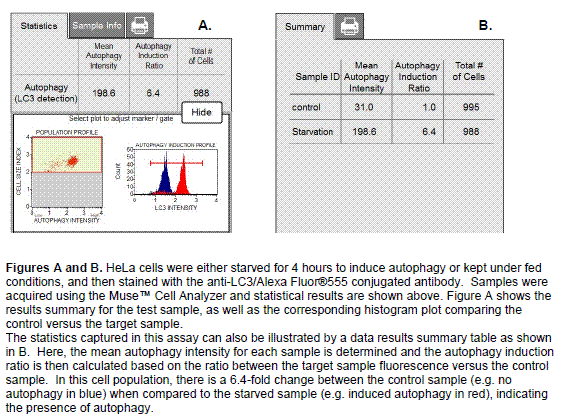
2) Since autophagy is a constitutive cellular degradation process, the use of an autophagy detection reagent (Autophagy Reagent A) will prevent the lysosomal degradation of LC3, allowing its quantification by flow cytometry. EMD Millipore’s Muse™ Autophagy LC3-antibody based kit includes an anti-LC3 mouse monoclonal antibody conjugated to Alexa Fluor®555, used to measure and track the levels of LC3 within the cell. The anti-LC3 Alexa Fluor®555 conjugated antibody and autophagy enabling reagents have been carefully evaluated to ensure optimal performance, alleviating the need for any additional validation of the kit reagents. This kit contains reagents A and B, along with an assay buffer to provide researchers a complete solution for autophagy analysis.The autophagy detection reagents and antibody have been optimized together to ensure the ability to measure and discriminate between cytosolic and lipidated LC3 to accurately measure the autophagic process. Data generated using the Muse™ Cell Analyzer along with the corresponding Muse™ software module provides statistical values measuring:
• Mean Autophagy Value (for both control and test samples)
• Autophagy Induction Ratio (Test sample fluorescence relative to control)
• Percentage of cells with increased autophagy (Test sample versus control)