

Bacteria can be analysed flow cytometrically when in suspension without fluorescent staining, numerous fluorescent dyes can be used to indicate the presence of bacteria, viability, DNA content and membrane potential.
Labelling bacteria
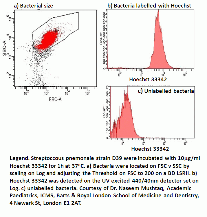
The presence of bacteria for image analysis or interactions with mammalian cells can be achieved by labelling with FITC, Bis-oxonol dye, DiBAC4(5) or Hoechst 33342.
Viability
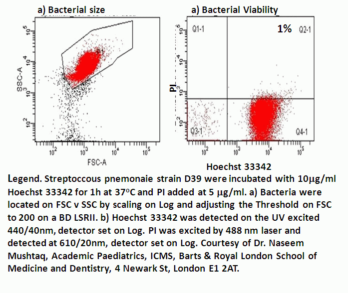
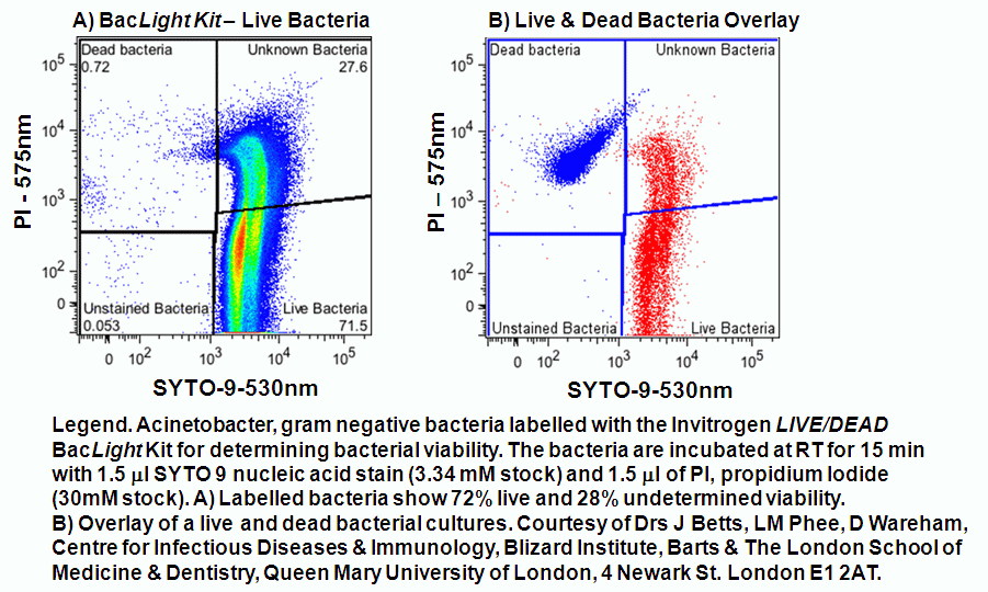
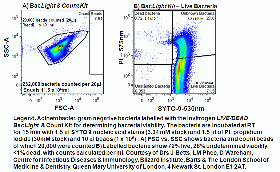
Bacterial viability can be assessed by commercial kits but DAPI and PI can be used to determine bacterial viability. This can be used in conjunction with Hoechst 33342. The Live/Dead BacLight kit from Invitrogen uses two nucleic acid dyes, PI to stain dead bacteria and SYTO9, a green nucleic acid stain which will only stain bacteria with an intact membrane. Gram negative and positive bacteria will take up these two dyes to differing degree's and so each species/strain of bacteria has to be titrated to show live (SYTO9+ve), dead (PI+ve) populations of bacteria, as well as a double stained population of unknown viability, see figure.
DNA content
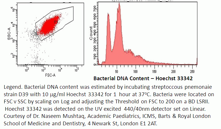
Bacterial DNA content can be assessed by the use of Hoechst 33342 or PI on fixed bacteria.
Membrane potential
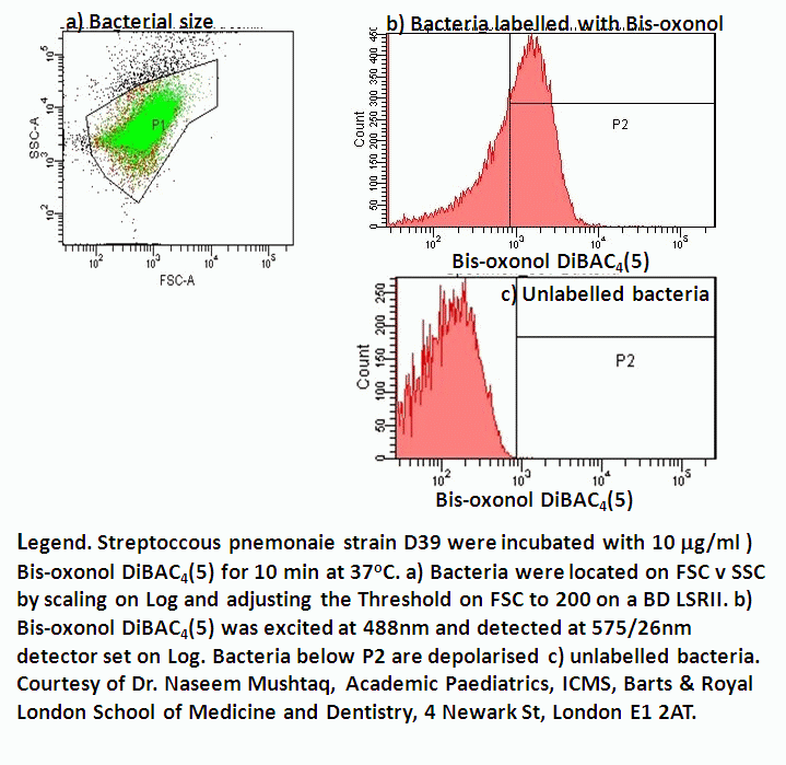
Measurement of bacterial membrane potential by Bis-oxonol is useful in combination with determination of bacterial viability giving a differential analysis.
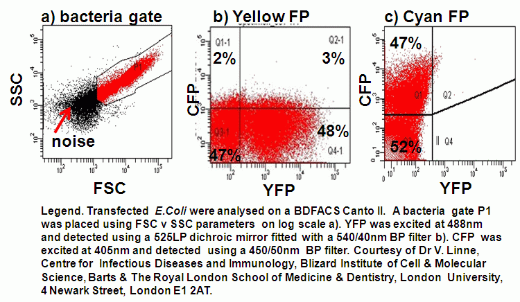
Fluorescent Protein (FP)
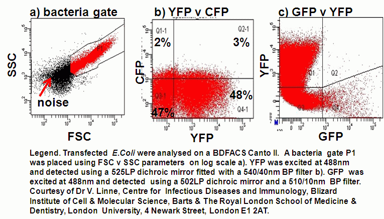
Bacteria can be transfected with FPs including Cyan or CFP,Green or GFP and Yellow or YFP that can be detected by flow cytometry.
Counting
Bacteria can also be counted using the Invitrogen BacLight kit which is also supplied with calibrated 6 mm microspheres at 100 million/ml. Thus 1 ml of the bead solution is also added to 1 ml of bacteria to enable the counting of bacteria per volume, see figure.
- Labelling protocol
- Viability protocol
- DNA content protocol
- Membrane potential protocol
- Live Dead BacLight Kit (L34856)
Publication.
VA Gant, G Warnes, I Phillips, GF Savidge. The application of flow cytometry to the study of bacterial responses to antibiotics. J Med Microbiol 39,1475-154, 1993.