

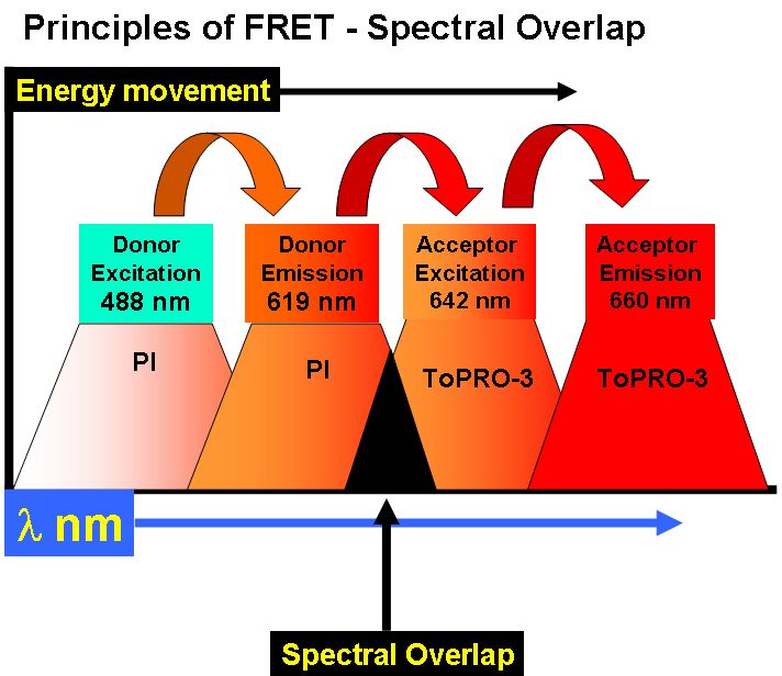
Fluorescence Resonance Energy Transfer or FRET can be used to show that molecules are within 10 nm of each other by nature of the transfer of energy via a long range dipole-dipole coupling mechanism. A fluorescent donor molecule is excited by a light source, the energy is non-radiatively transferred to an acceptor molecule, see figure Principles of FRET.
The efficiency of this energy transfer depends upon the distance between acceptor and donor, the degree of spectral overlap between the emission spectra of the donor and absorption of spectra of the acceptor and the relative parallel orientation of donor and acceptor dipoles. The distance at which the energy transfer is 50% efficient is called the Forster Radius (Ro). The magnitude of the Forster Radius for each fluorophore is also dependent upon the relative quantum yield of the fluorophore.
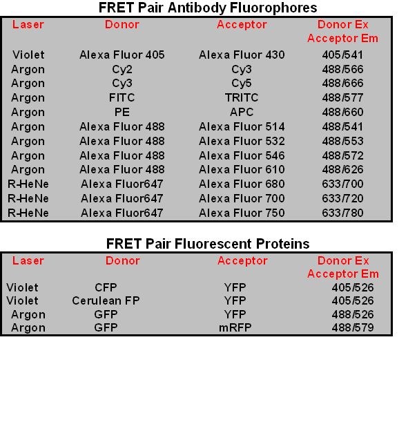
FRET Pairs
Thus the choice of fluorophores for a FRET experiment, or FRET pairs of donor and acceptor molecules is important given the range of spectral properties of fluorophores. Originally FITC & TRITC was used as FRET pairs. More recently the Cy dyes have been used as FRET pairs e.g. Cy2-Cy3 or Cy3-Cy5. The Alexa Fluor Dyes also act as FRET pairs e.g. Alexa Fluor 488 and Alexa Fluor 546. Fluorescent Proteins such as CFP-YFP, GFP-YFP and GFP-mRFP also act as FRET pairs, see table for more detailed information.
FRET can used to measure BrdU incorporation by the use of DNA dyes PI and ToPro-3.
FRET based gene reporter systems
A gene reporter system developed by Invitrogen uses FRET-based fluorescent substrates, CCF2 and CCF4.
Fluorescent Protein based FRET
Fluorescent proteins (FPs) can also be used to detect protein interactions or biological processes such as phosphorylation. The most commonly used FRET pairs of FPs are CFP-YFP, but GFP-YFP and GFP with DsRED, mRFP or RFP can also be used in flow cytometric detection of FRET. The development of gene transfection for single proteins expressing CFP and YFP at terminal ends of the protein allow FRET to occur when the protein undergoes a conformational change when acted upon by a specific biological process e.g. phosphorylation. CFP-YFP FRET can be detected by the Core Facilities LSR II analyser and Aria cell sorter enabling the sorting of FRET expressing cells.
Cells transfected with Fluorescent proteins (FPs) with the most commonly used FRET pairs are CFP-YFP, but GFP-RFP can also be used in flow cytometric detection of FRET. Such cultures of cells used for imaging of FRET can have varying levels of cells displaying FRET. In an example shown here cells were sorted according to high levels of CFP and YFP which showed a FRET level of 26% compared to the starting culture with 10% FRET. Imaging of FRET by the Sensitization method was made considerably easier by pre-sorting cells expressing higher levels of FRET.
Lipophillic carbocyanine dyes DiO and DiI act as donor and acceptor FRET pairs. This phenomena can be used to detect fused cells when the two original cell types are previously labelled with DiO and DiI. The enhanced DiI signal allows the detection of fused hybridoma cells.
The dimer constituents of receptors can also be determined be they homodimers or hetrodimers by FRET. In the case of homodimer receptors, antibodies against for example CD8 alpha-alpha, the same antibody clone is used with two FRET paired fluorophores e.g. Pacific Blue and Alex Fluor 488. Simultaneous labelling with the two antibodies, Pacific Blue acting as a Donor and Alexa Flour 488 as the Acceptor allow the detection of any FRET in the Violet laser 530nm channel in this case detecting CD8 alpha-alpha homodimer receptors. Hetrodimer receptors can also detected in a similar manner.
Antibodies conjugated with Alexa Fluor dyes such as Alexa Fluor 488 and for example 555 can also act as donor and acceptor fluorophores to detect FRET. Such Alexa Fluor labelling of 3-Phosphoinositide dependent protein kinase 1 (PDK1) and phospholipase Cg1(PLCg1) on fixed cells allows FRET to measured in the 575nm argon laser channel (PE) dynamically over time enabling easy and meaningful display of the FRET signal due to PDK1 interaction with PLCg1. EGF stimulation of transiently transfected breast epithelial MDA-MB-231 cells can show that protein kinases such as PDK1 regulate phospholipase C, PLCg1 which has been implicated in cancer cell invasion.
FRET Publications
C Raimondi, A Chikh, AP Wheeler, T Maffucci, M Falasca. A novel regulatory mechanism links PLCg1 to PDK1. J Cell Sci Epub May18 2012.
G Warnes. Flow Cytometric Applications of FRET - Fluorescence Resonance Energy Transfer. The Biomedical Scientist, 53(2) p119-121,Feb 2009.
A Vossenkamper, O Marches, PD Fairclough, G Warnes, AJ Stagg, JO Lindsay, PC Evans,LA Luong, NM Croft, S Naik, G Frankel, TT McDonald. Inhibition of NF-kB signaling in human dendritic cells the enterpathogenic Escherichia coli effector protein NIeE . J Immunol 185: 4118-4127, 2010.