
Apoptosis can be defined as 'gene-directed cellular self-destruction'; it is sometimes referred to as 'programmed cell death' or Type I PCD, although this is really a phenomenon where cells are programmed to die at a particular point, e.g. during embryonic development, and even here cells may go through an apoptotic pathway. However, 'classic' apoptosis is certainly a distinct process from other forms of oncosis leading to necrosis. The new term 'Cell Necrobiology' is an encompassing term for all modes of cell and can be defined as "various modes of cell death: the biological changes, which predisose, precede and accompany cell death; as well as the consequences and tissue response to cell death". The term necrobiology thus includes all the many forms of apoptosis besides 'classic' PCD, including caspase independent apoptosis-like PCD, autophagy or Type II PCD, mitotic catastrophe, necrosis-like PCD, necrosis or oncosis defined as accidental cell death and cell senescence.
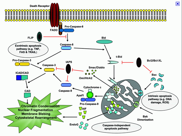
Apoptosis can be activated by the 'extrinsic pathway' which is mediated via the Cell Death Receptor pathway. This pathway is typically activated by TNF binding to the receptor, see model of the extrinsic and intrinsic pathways below:-
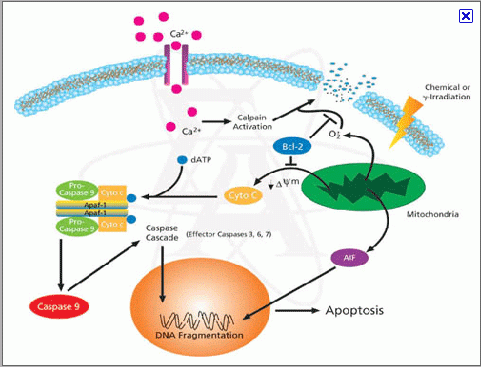
The 'Intrinsic pathway' can be drug induced, UV-B irradiation, g-irradiation, and reactive oxygen and is mediated via mitochondrial responses to such cellular insults, see model below:-
Classic apoptotic cells can be recognized by a characteristic pattern of morphological, biochemical and molecular changes, which may be broadly and chronologically defined as:
Morphological Changes- Cell shrinkage
- Cell shape change
- Condensation of cytoplasm
- Nuclear envelope changes
- Nuclear fragmentation
- Loss of cell surface structures
- Apoptotic bodies
- Cell detachment
- Phagocytosis of remains
- Free calcium ion rise
- bcl2/BAX interaction
- Cell dehydration
- Loss of mitochondrial membrane potential
- Proteolysis
- Phosphatidylserine externalisation
- Formation of Mitochondrial Permeability Transition Pore
- Lamin B proteolysis
- DNA denaturation
- 50-300kb cleavage
- Intranucleosomal cleavage
- Protein cross-linking
Many of these changes may be measured by flow cytometry and some are described below.
Publications.
A Vossenkamper, G Warnes. Thapsigargin induced apoptosis occurs with a delayed DNA damage response, Br J Cancer Res 2(4):314-319, 2019. (in press ).
A Vossenkamper, G Warnes. Flow cytometry reveals the nature of oncotic cells, Int. J. Mol. Sci. 20(18), 4379; 2019.
D Bergamaschi, A Vossenkaemper, WYJ Lee, P Wang, E Bochukova, G Warnes. Simultaneous polchromatic flow cytometric detection of multiple forms of regulated cell death. Apoptosis, 2019. (DOI: 10.1007/s10495-019-01528-w)
A Popat, AA Patel, G Warnes. A flow cytometric study of ER Stress and autophagy, Cytometry A, DOI: 10.1002/cyto.a.23665, 2018.
A Vossenkamper, G Warnes. A flow cytometric immunophenotyping approach to the detection of regulated cell death processes. Journal of Immunological Sciences, 2 (5) 2018.
HL Lee, R Pike, MHA Chong, A Vossenkamper, G Warnes. Simultaneous flow cytometric immunophenotyping of necroptosis, apoptosis and RIP1 dependent apoptosis Methods, 1, 134-135, 56-66, 2018.
Luo J, Lee S, Wu D, Yeh J, Ellamushi H, Wheeler AP, Warnes G, Zhang Y, Bo X. P2X7 purinoceptors contribute to the death of Schwann cells transplanted into the spinal cord. Cell Death Dis. Oct 3;4:e829. doi: 10.1038/cddis.2013.343, Oct 2013.
TS MacFie, R Poulsom, A Parker, G Warnes, TBoitsova, A Nijhuis, N Suraweera, APoehlmann, JSzary, R Feakins, R Jeffery, RW Harper, AM Jubb, JO Lindsay, A Silver. DUOX2 and DUOXA2 form the predominant enzyme system capable of producing the reactive oxygen species H2O2 in active ulcerative colitis and are modulated by 5-aminosalicylic acid. Inflammatory Bowel Disease, Jan 2014.
SN Jayasinghe, G Warnes, CJ Scotton. Bio-electrosprayed living microenvironments implanted into mouse models. Macromolecular Bioscience 11(10), 1364-69, 2011.
SM Janes, TA Ofstad, DH Campbell, A Eddaoudi, G Warnes, D Davies, F Watt. PI3-kinase dependent activation of apoptotic machinery occurs on commitment of epidermal keratinocytes to terminal differentiation. Cell Res 19(3): 328-339, 2009.