
Cell proliferation
Cell proliferation can now be measured flow cytometrically by the use of numerous probes including, Carboxyfluorescein Diacetate, Succinimidyl Ester or CFSE, PHK26, and now Violet Cell Trace, enabling researchers to easily monitor the proliferation rate of cells transfected with GFP, the original CFSE being in the same channel as GFP. If violet laser diodes are not available, a Red HeNe diode can be used to measure cell proliferation by the use of Cell Trace Far Red DDAO-SE.
Carboxyfluorescein Diacetate, Succinimidyl Ester (CFDA, SE)
Cell proliferation may be assessed by flow cytometry. Cell are labelled with the dye CFDA,SE which spontaneously and irreversibly couples to both intracellular and cell surface proteins. When cells divide CFDA,SE labelling is distributed equally between the two daughter cells, which therefore have half the fluorescence of the parents. As a result, each successive generation in a population is marked by a halving of the cellular fluorescence intensity which is readily followed by flow cytometry.
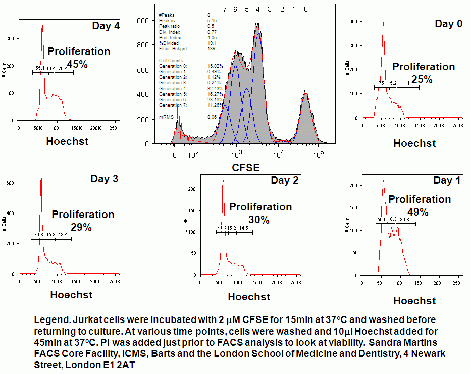
CFSE proliferation studies can be combined with live cell cycle analysis throughout a time course study of several days. This can be accomplished by incubating daily aliquots of CFSE loaded cells with 10 ug/ml of Hoechst 33342. Jurkat T-cells were studied for cell proliferation and live cell cycle analysis over a 5 day period, the cell cycle analysis shows that the number of proliferating cells changes over the 5 day period, see figure.
- Tracking of asynchronous cell division using CFDA,SE or CFSE
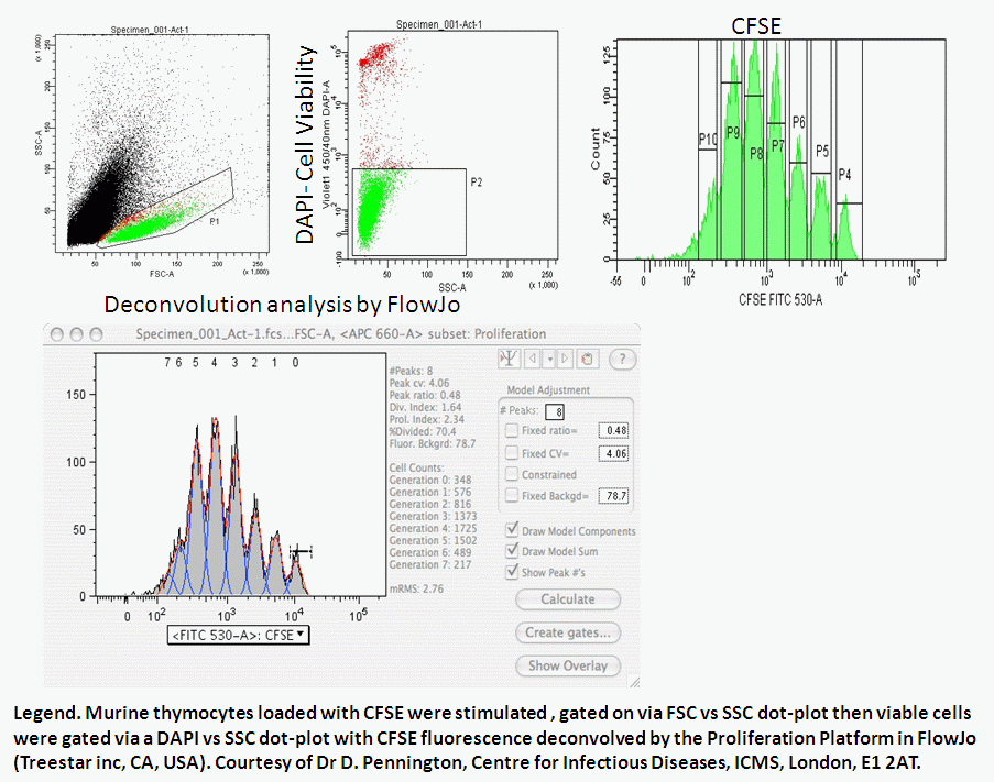
- Mouse thymocyte proliferation
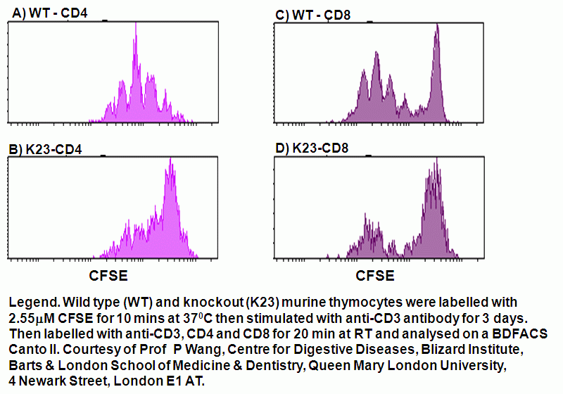
- CD4-CD8 mouse thymocyte anti-CD3 stimulation
- Murine thymocyte CD25-CSFE profiling
- Immunophenotyping and CFSE labelling
- T-cell proliferation studies combined with cell cycle analysis
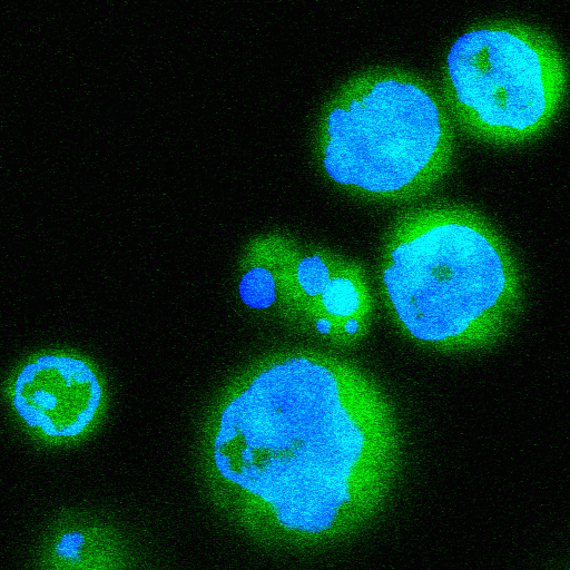
- Image of Jurkat cells loaded with CFSE and Hoechst 33342
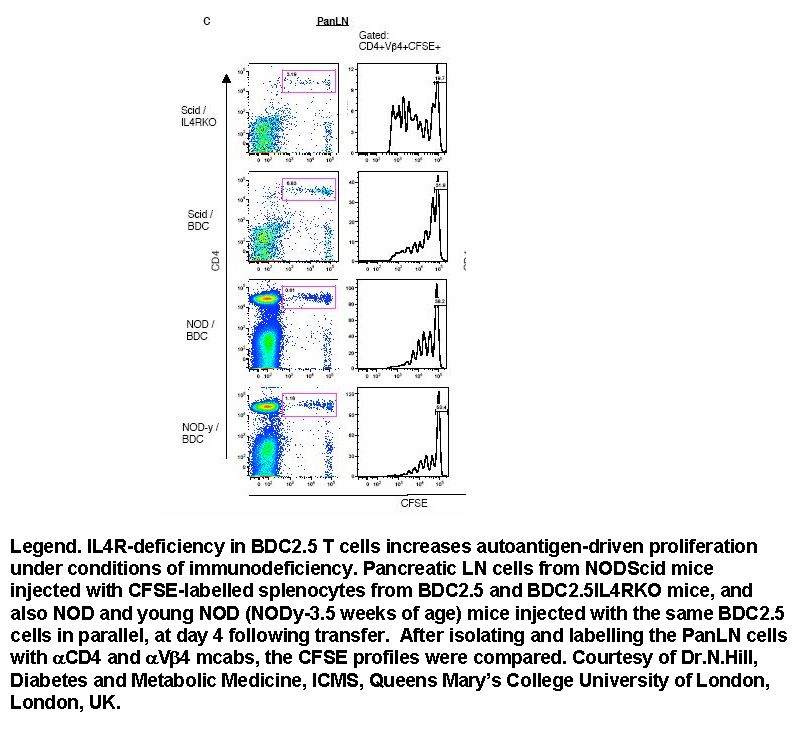
The effect of lymphopenia upon antigen-driven proliferation of auto-reactive CD4 T-cells (or CD4 +ve VB4+ve) was investigated monitoring proliferation with CFSE in SCID/IL4-R KO mice.
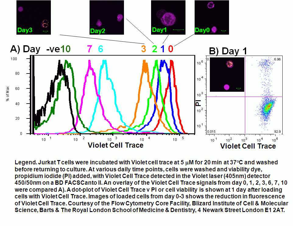
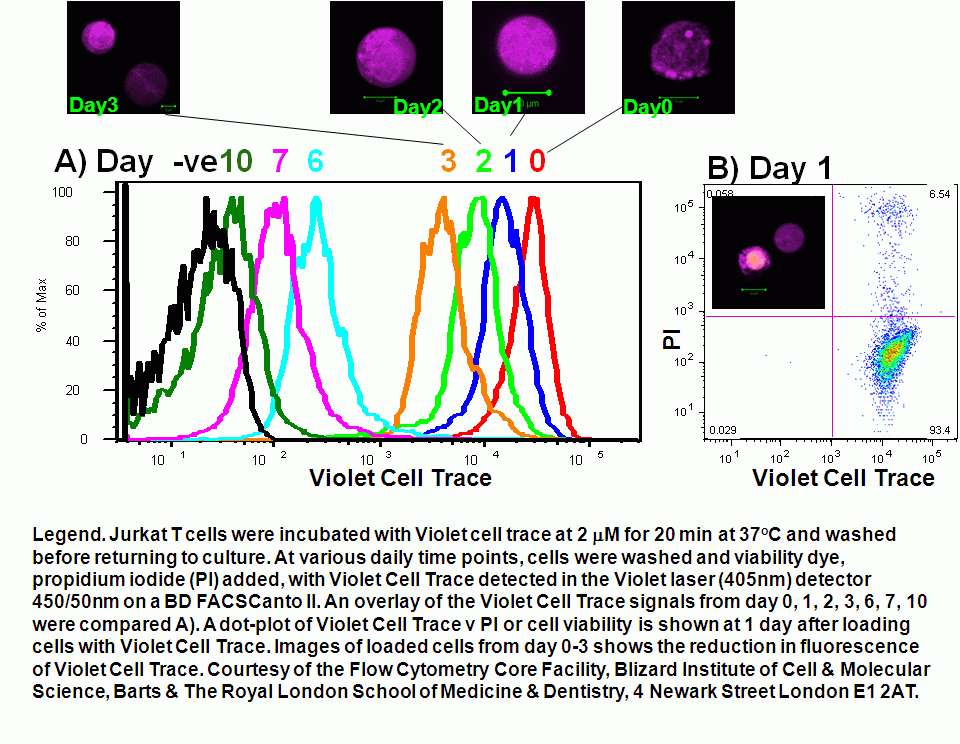
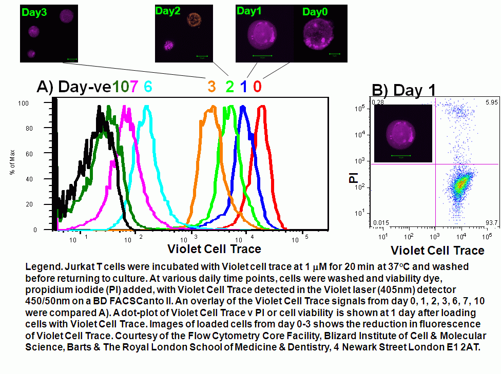
Violet Cell Trace
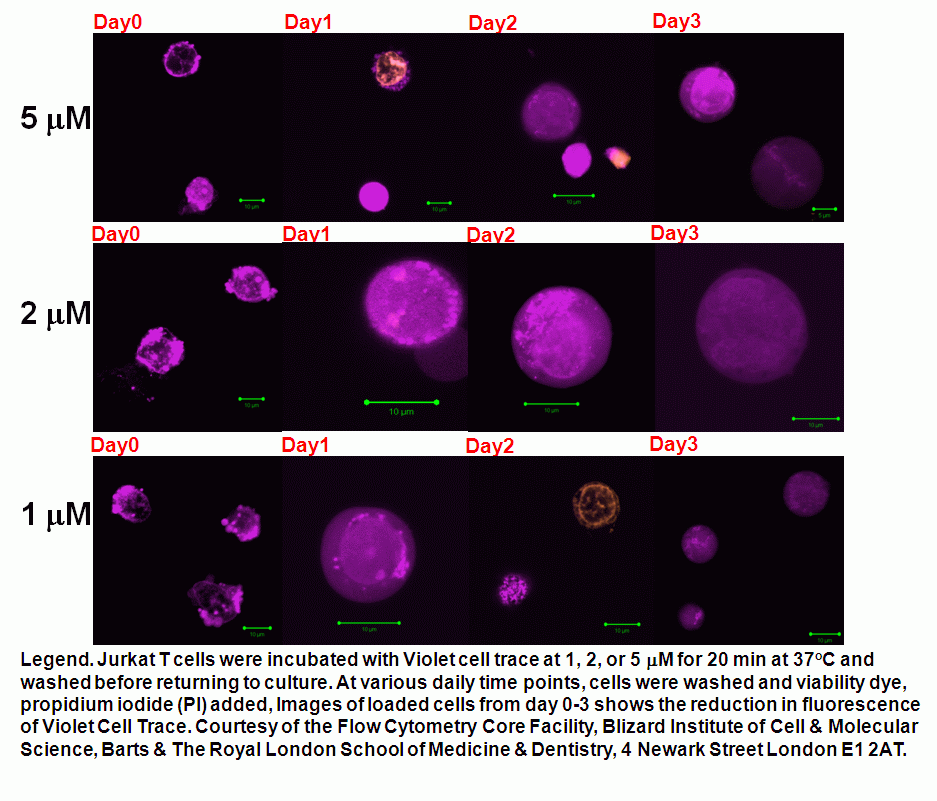
This new probe, Violet Cell Trace is easily loaded into cell lines such as Jurkat T-cells which can be monitored for proliferation for up to 10 days and be easily imaged by confocal microscopy, although at high concentrations this probe can bleed into the GFP channel. Jurkat cells loaded with 5, 2 or 1mM Cell Trace Violet were compared over a 10 day incubation period with fluorescence intensities compared for the 3 concentrations, see figures for 1, 2, 5mM. Protocol
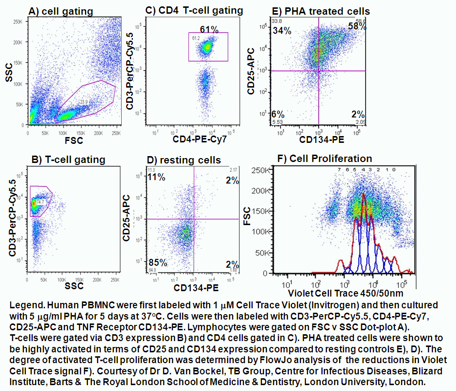
Human PBMNC lymphocytes were pre-labeled with 1 mM Violet Cell Trace for 20 mins at 37C.Lymphocytes were then PHA activated for 5 days. Activated and resting lymphocytes were then surface immunophenotyped for CD3-PerCP-Cy5.5, CD4-PE-Cy7 and activation markers CD25-APC (IL-2 receptor) and CD134-PE (TNF receptor). Activated lymphocytes were then analysed for cell proliferation by measuring the reduction in Violet Cell trace (450/50nm channel) signal by computer analysis using FlowJo software, see figure.