
G Warnes Live cell analysis of necroptosis 2018.
G Warnes Immunophentyping and modulation of oncotic repsonse, 2018.
G Warnes. Flow cytometric immunophenotyping of necroptosis by RIP3, MLKL and Caspase-3 analysis, 2018.
A Vossenkamper, G Warnes. Flow cytometric analysis of necroptosis, RIP1 dependent apoptosis, apoptosis, autophagy, ER stress, parthanatos, DNA Damage. 2017-8.
HL Lee, R pike, MHA Chon, A Vossenkamper, G Warnes. Flow cytometric analysis of necroptosis, RIP1 dependent apoptosis and apoptosis. 2016-7.
A Popat, G Warnes. An investigation into the the links between ER Stress, autophagy, apoptosis and necroptosis. 2016.
A A Patel, BSc project. An Investigation into ER stress induced Autophagy. September 2014.
T T Pham, BSc project., An investigation into autophagy and cell cycle changes during nutrient starvation. September 2013.
R Akinlade, Neuroscience MSc project. Lithium in Multiple Sclerosis: Effects on cell survival mechanisms? June 2013.
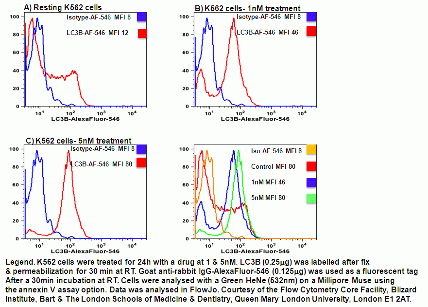
G Warnes. Imaging of LC3B-AF-546 and Muse Analysis. June 2013.
E James, Neuroscience project. March 2013.
O Dadoun, BSc project. An Investigation into Autophagy During Nutrient Starvation. September 2012.
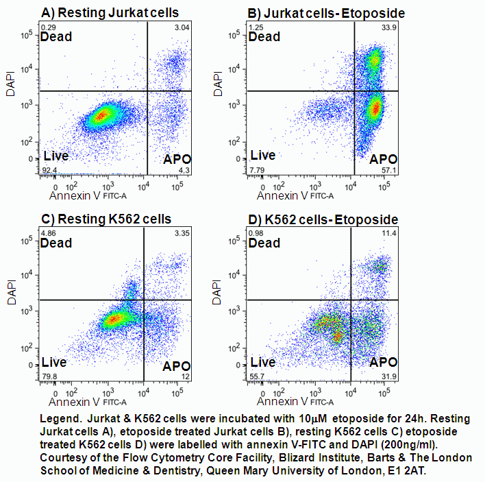
B Wilbourn. Etoposide Induction of Apoptosis. September 2012
G Warnes. NADH detection. October 2011.
N Panchal, BSc project. Developing a New Flow Cytometry Diagnostic Technique Using LysosTracker Dyes to Detect Autophagy. September 2011.
S Chikte BSc project. LysoTracker Green Analysis Using Flow Cytometry to Study Autophagy. September 2011.
G Warnes. Real Time Flow Cytometry for the Kinetic Analysis of Oncosis. July 2010.
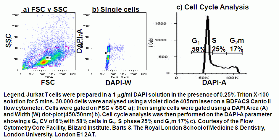
G Warnes. Cell Cycle Analysis:- Triton X-100 cell treatment for violet laser excitation of DAPI. May 2010.
N Mushtag. Bis-oxonal loading of bacteria 2009.
