
Type II Apoptosis
Macroautophagy or autophagy translates as 'self eating' which is an apoptotic process in response to nutrient starvation, protein aggregation, Endoplasmic Reticulum (ER) stress, calcium overload, hypoxia, removal of damaged organelles and proteasome impairment. Autophagy can be triggered as a cell survival mechanism and hence may prevent cell death and thus it is integral to human health in terms of organism development, cell homeostasis and is involved in a wide range of diseases including cancer, neurodegeneration, aging and the innate immune response to pathogens.
Autophagy was first fully described in 1963 by Christian de Duve who observed cytoplasmic material such as mitochondria and endoplasmic reticulum being engulfed by double-membrane vesicles or autophagosomes and after fusion with lysosomes digestion by lysosomal enzymes in the newly formed auto-lysosome.
Autophagy can be characterized morphologically in a similar manner to that of other forms of cell death by cell shrinkage, formation of autophagosomes and degradation of cell organelles. There are numerous signalling routes that regulate autophagy, these include mTOR and JNK signalling. Autophagy can be induced by nutrient starvation, formation of cytosolic protein aggregates, ER stress, calcium overload and damaged organelles.
There are numerous methods of inducing autophagy including serum and total nutrient starvation and rapamycin which both inhibit mTOR signalling. Chloroquine can also induce autophagy as well as ER-ATPase inhibitor, Thapsigargin which causes autophagy of the ER.
Morphological Changes
- Cell shrinkage
- Double membrane vesicles or autophagosomes
- Degradation of organelles
- mTOR
- PI3 kinases
- UPR stress sensors
- Kinases - JNK
- Bcl-2
- IP3-receptor
Stimulus
- Nutrient starvation
- Protein aggregation
- Ischemia
- Hypoxia
- Calcium overload
- ER Stress
- Damaged organelles
- Proteasome impairment
Model of Autophagy
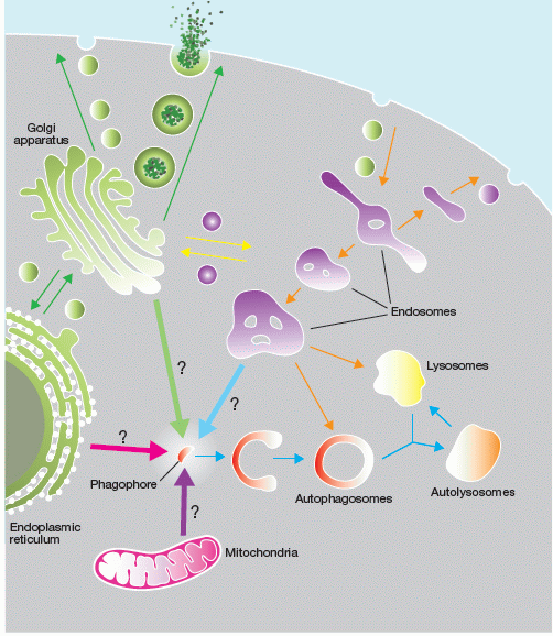
Figure by SA Tooze, The orgin of the autophagosomal membrane, Nature Cell Biology, 12, (9), p831-835, 2010.
Publications.
S Chikte, N Panchal, G Warnes. Use of LysoTracker dyes: A flow cytometric study of autophagy. (manuscript submitted for review) 2013.
N Panchal, S Chikte, BR Wilbourn, UC Meier, G Warnes. Flow cytometric measurement of cell organelle autophagy: Autophagy, InTech Open Access Publisher ISBN 980-953-307-971-9, 2013
A Rosello, G Warnes, UC Meir. Cell death pathways and autophagy in the central nervous system and its involvement in neurodegeneration, immunity and CNS infection: to die or not to die - that is the question. Clinical & Experimental Immunology Apr, 162(1), 52-7, 2012.