
Role of microtubule protein LC3 in autophagy
There are numerous methods of inducing autophagy including serum starvation and rapamycin which both inhibit mTOR signalling. Chloroquine interestingly induces the formation of autophagosomes but blocks the formation of autophagolysosomes and hence ultimately is an inhibitor of autophagy. The ER-ATPase inhibitor, Thapsigargin has also been reported to induce autophagy of the ER.
Acridine Orange (AO) which is commonly employed to measure DNA and RNA levels within cells can also be employed to measure the level of acidic granule formation within cells undergoing autophagy, which show an increase in the red signal.
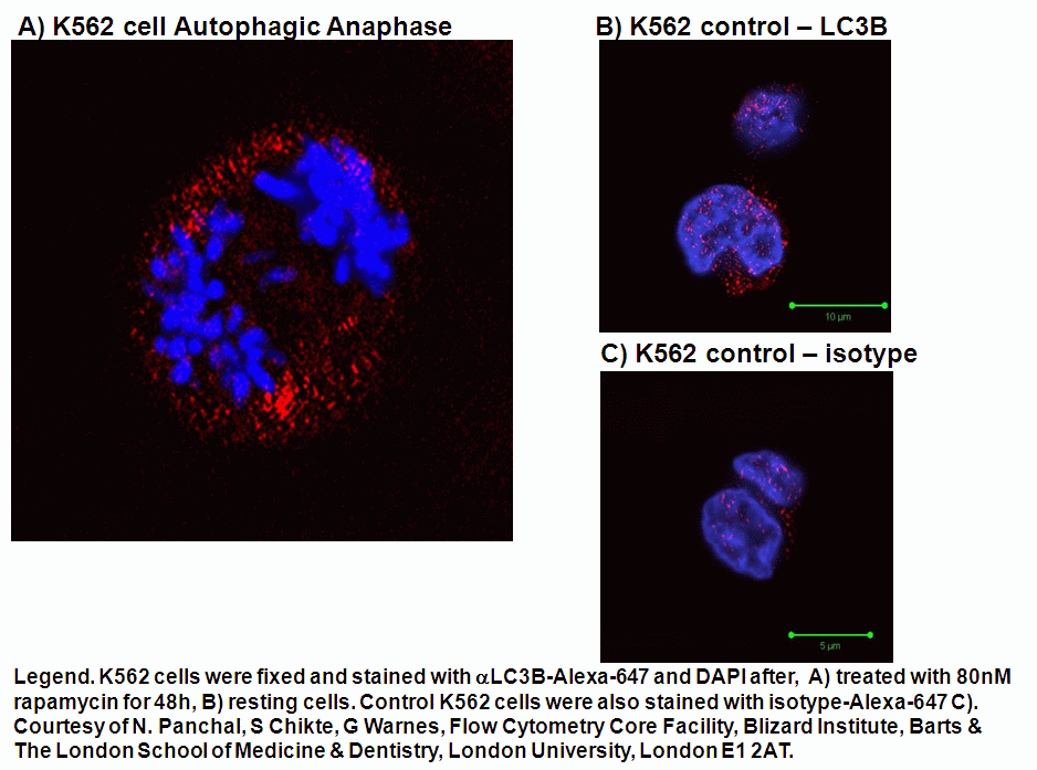
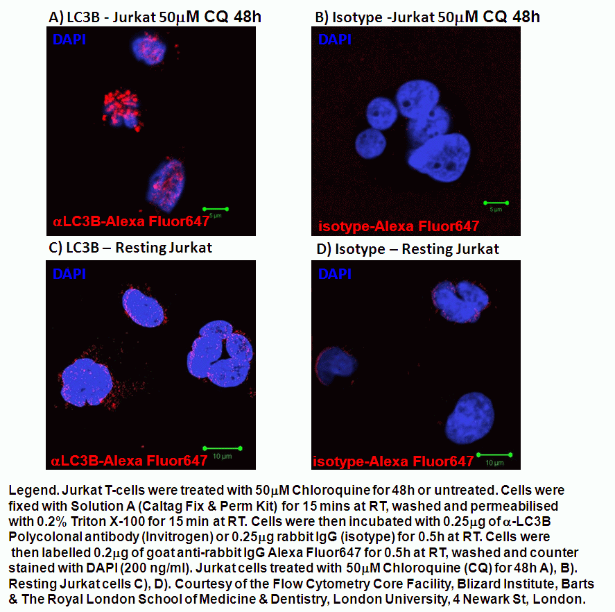
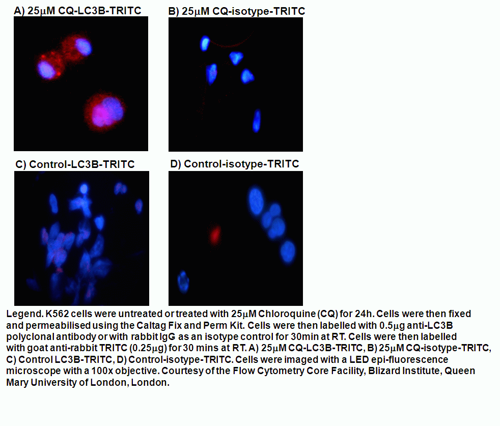
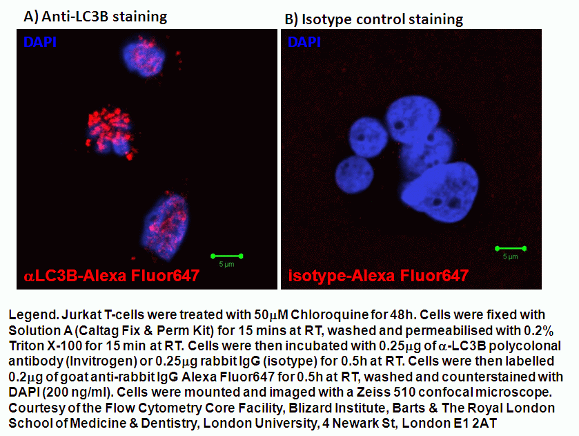
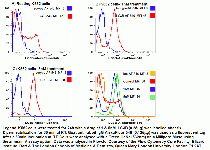
The current methods of determining the presence of autophagy is based on the fact the microtubule protein, LC3 normally present in the cytoplasm. During the autophagic process or flux LC3 is cleaved and lipidated with the phospholipid, phosphatidyl-ethanolamine (PE) to form LC3B in autophagosomes, see figure. After fusion of the autophagosome with lysosomes, to form the single membrane bound, autolysosome the the internal LC3B is reduced. The LC3B can be tagged with GFP (fluorescence is acid sensitive) and RFP (acid resistant) and imaged by fluorescence microscopy or analysed flow cytometrically. The LC3B levels can also be measured by the use of polyclonal LC3B antibody tagged in this case with Alexa Fluor 647, which can then be analysed flow cytometrically. LC3B can also be tagged with Alexa Fluor 546 and analysed using the Green HeNe laser on a Millipore Muse instrument using the Annexin V assay program, see figure.
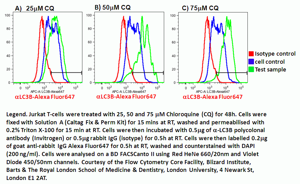
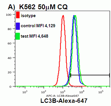
Chloroquine, an anti-malarial drug not only induces autophagy but inhibits the fusion of lysosomes with the autophagosomes to form autolysosomes. Thus chloroquine causes a build up of autophagosomes and thus a large LC3B signal, which is thus akin to the use of Golgi stop agents Brefeldin A and monensin, the increased signal can be easily detected flow cytometrically after 48 hours treatment with 25, 50, 75mM chloroquine, see figure. Similarly K562 erythromyeloid leukeamia cells can be treated with chloroquine to induce autophagy.
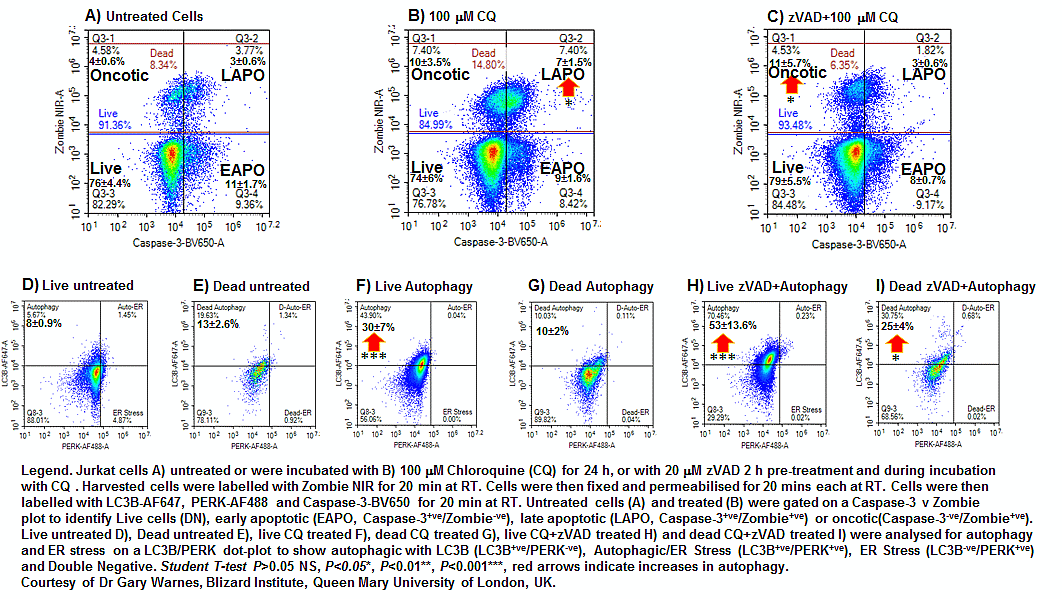
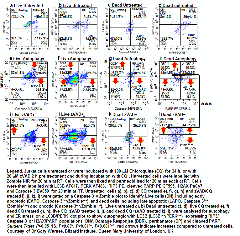
Autophagy initiated by chloroquine (CQ) is often associated with ER stress and ultimately cell death by apoptosis. These can be all measured in cells by the use of a fixable viability dye, and antibodies against intracellular caspase-3 to measure apoptosis, LC3B to measure autophagy and PERK to measure ER stress, see figure. Blockade of apoptosis with pan-caspase blocker, zVAD blocks CQ induced apoptosis but up-regulates autophagy in both live and dead cells, see figure.
Autophagic cells induced by CQ also show differing degrees of resting (RIP3+ve/Caspase-3-ve), early apoptotic (Zombie-ve/RIP3-ve/Caspase-3+ve), late apoptotic (Zombie+ve/RIP3-ve/Caspase-3+ve)and live and dead RIP1-dependent apoptosis (Zombie+/--ve/RIP3+ve/Caspase-3+ve). These autophagic cells also showed less DNA Damage (H2AX+ve/PARP-ve) when undergoing autophagy while pre-treatment with zVAD removed this affect of autophagy, see figure. While dead autophagic cells showed increased levels of DNA Damage, see figure. Parthanatos or hyper-activated PARP (H2AX+ve/PARP+ve) and cleaved PARP (H2AX-ve/PARP+ve) was also detected in these live and dead autophagic cells to differing degrees, see figure.
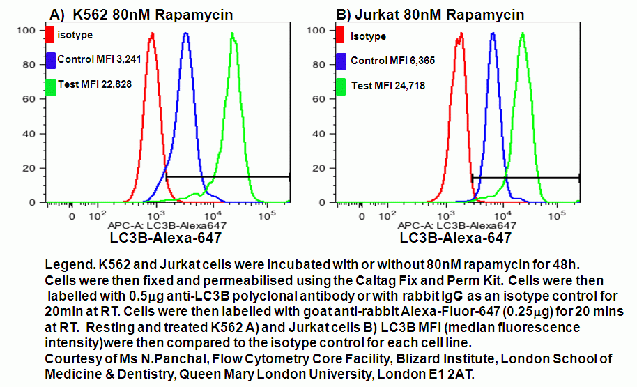
Rapamycin induced autophagy can also be detected flow cytometrically by the use of anti-LC3B in Jurkat and K562 cells even though there is no autophagosome stop activity, see figure.
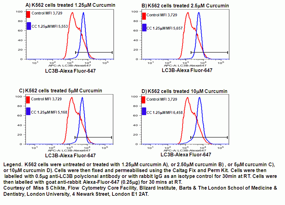
The Indian spice, Tumeric or Curcumin also induces autophagy as well as apoptosis the increase in autophagosomes can also be measured flow cytometrically by the use of LC3B, see figure. LC3B reveals that curcumin induces a dose response in K562 cells.