
Reticluophagy
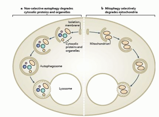
The process of autophagy is a cell survival mechanism that occurs when the cell is under stress, via external environmental pressures, including the lack of nutrients, or via the internal microenvironment of the cell, including the replacement of old and defective organelles such as mitochondria.
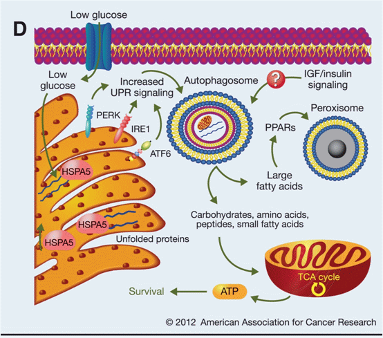
Autophagy is also induced by the formation and collection of mis-folded proteins in the ER, which causes ER-stress within the cell. Limited autophagy of the cell allows the generation of ATP from digested organelles leading to its ultimate survival. Whilst prolonged adverse conditions results in the death of the cell by the autophagic process.
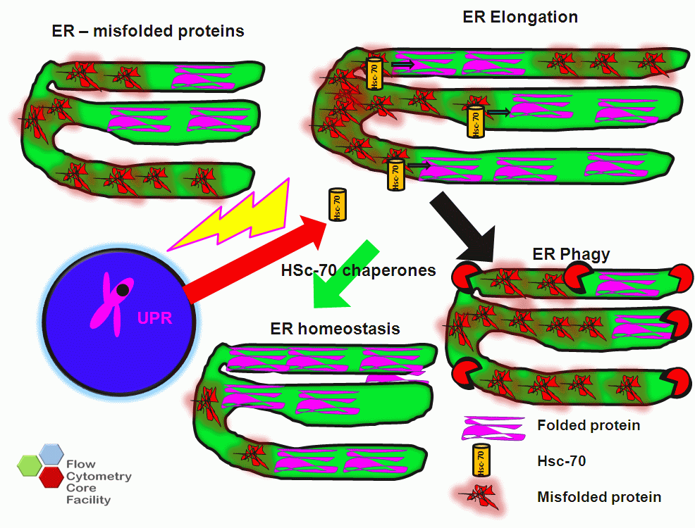
The ER is responsible for the folding and then delivery of proteins via the secretory pathway to a functional site. Mis folded proteins accumulate in the lumen of the ER due to high protein folding demand on the ER. Only properly folded proteins are secreted with maintenance of the plasma membrane structure and ER folding capacity being under ER homeostatic control. Once a threshold of mis-folded protein accumulation has been reached, a signal activates the ER to nucleus signalling pathway or the unfolded protein response (UPR) causing ER chaperone proteins to be synthesised, which refold the mis folded proteins and translocate these proteins to the cytoplasm for degradation by the proteasome. This process results in an increase of the ER capacity to fold proteins and maintain ER homeostasis. This increase in ER capacity is physically achieved by an increase in size of the ER at early stages of autophagy, see figure. The process of reticulophagy is also cell cycle dependent with the loading of cell permeant DNA dye, Hoechst 33342 can be done at the same time as loading ER Tracker Red, see figure.
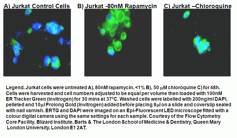
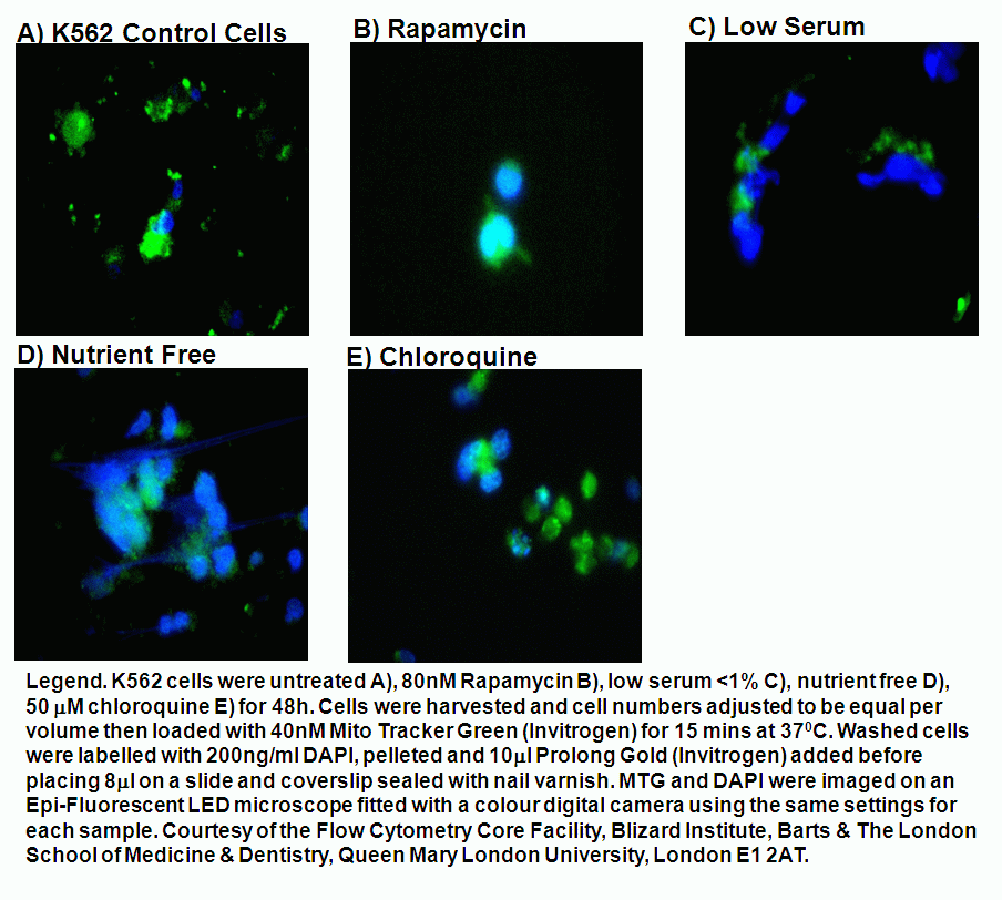
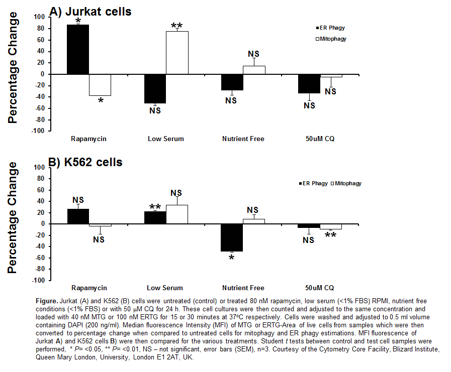
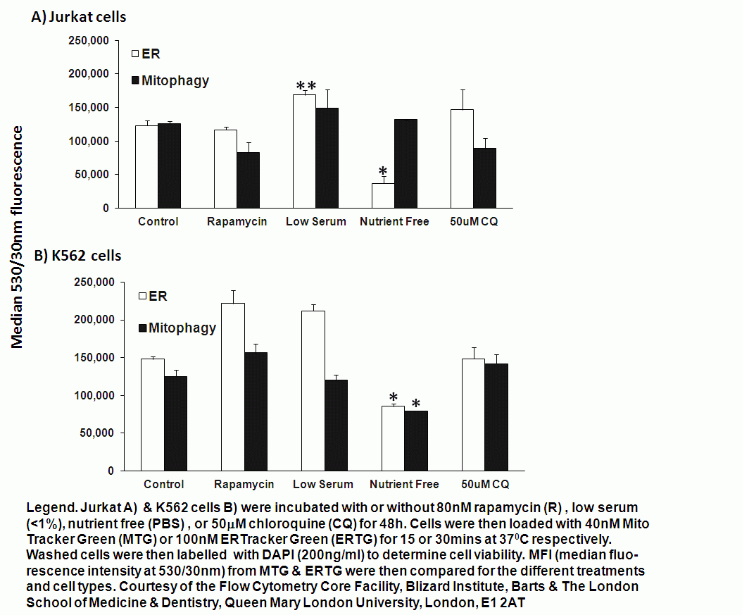
If the protein folding demand continues to increase, this will ultimately result in the phagy of the ER itself, which can be caused by ER stress (induced in vitro by tunicamycin and dithiothreitol, DTT), and also multiple mechanisms that induce autophagy e.g. drugs such as rapamycin and nutrient starvation, see figure.
Schematic of Reticulophagy
Mitophagy
Microautophagy or mitophagy is another an autophagic process, which is an organelle specific process, whereby dysfunctional mitochondria are removed from the cell by this autophagic response to reduce the number of reactive oxygen species (ROS) over-producing mitochondria. These malfunctioning mitochondria can be aged or those with a high level of mutations in the mtDNA induced by high levels of ROS. Mitochondrial DNA has 10-20 times more mutations than nuclear DNA.
Mitochondria not only produce ATP from the process of oxidative phosphorylation on the inner mitochondrial membrane but also reproduce by the process of fission. This produces two daughter mitochondria one of which hosts the damaged parts with reduced inner membrane potential of the original mitochondria but also a fully functional daughter. The fission process can also stop excessive enlargement of mitochondria. Whilst, mitochondrial fusion of damaged mitochondria dilutes the individual mitochondrion level of damaged macromolecules, this also prevents the early removal of such mitochondria.
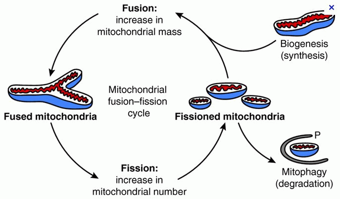
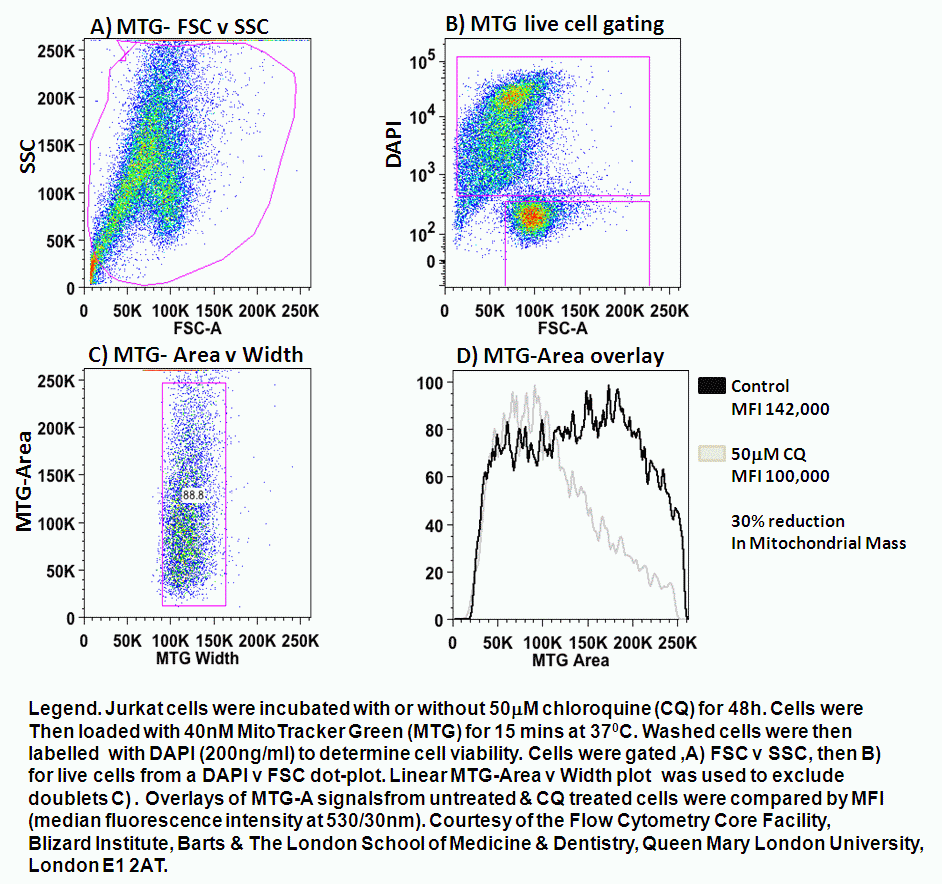
Mitophagy is a physiological process, which occurs during erythrocyte differentiation and during nutrient starvation, see figure. The level of relative mitochondrial fusion and fission within cells maintains mitochondrial homeostasis. Thus mitophagy provides a mechanism by which aged or ROS damaged mitochondria are ultimately removed resulting in the survival of the cell in question.
