
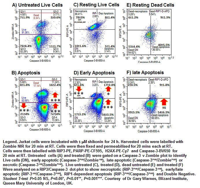
Classic apoptosis with activation of caspase-3 in the absence of RIP3 in cells without or with a permeable plasma membrane identifies early and late apoptosis respectively. Pre-labelling of cells with a fixable cell viability dye and subsequent fixing and permeabilisation allows intracellular labelling of active caspase-3 and RIP3 to identify these two stages of classic apoptosis. This is refinement of the now classic annexin V binding assay which although reasonably cheap and easy to use is not specific for any form of Regulated Cell Death (RCD).
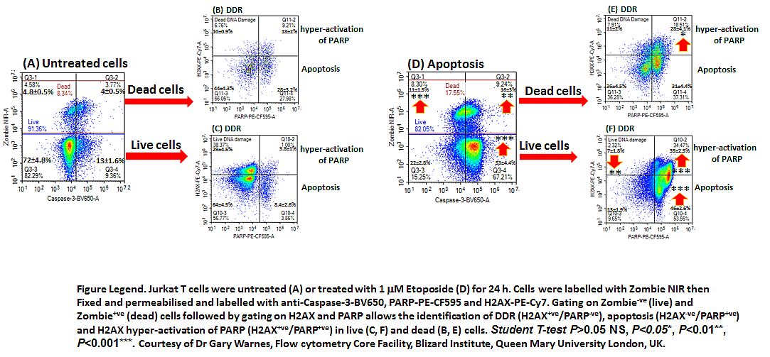
Etoposide induces apoptosis which can be immunophenotyped using a fixable live-dead probe along with Caspase-3 and RIP3 as described above. Further immuno phenotyping with H2AX and cleaved PARP permits the identification of DNA Damage (H2AX+ve), apoptosis (PARP+ve), H2AX hyper-activation of PARP (H2AX+ve/ PARP+ve) or parthantos and Double Negative (DN) cells, see figure.