
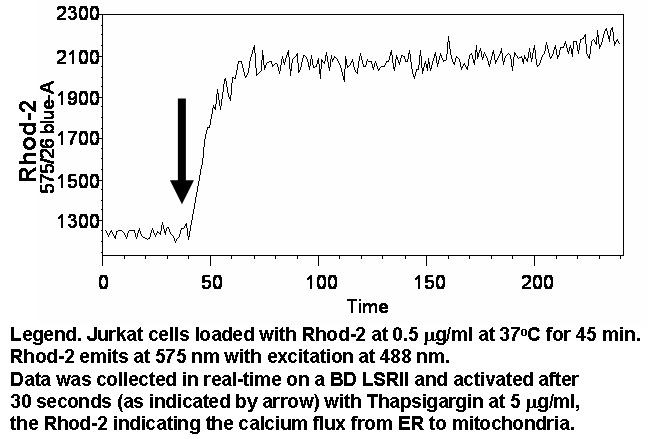
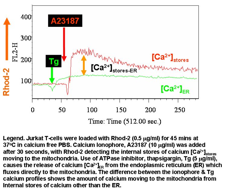
Rhod-2-AM when loaded into cells locates mainly to the mitochondria. Thapsigargin (Tg), an inhibitor of ER ATPase whose action releases internal calcium stores from the ER which then traffick's directly to the mitochondria. Thus Rhod-2 can be used to indirectly measure the amount of calcium in the ER. The release of all internal calcium stores within the cell by ionomycin a calcium ionophore, can be used to determine the amount of calcium fluxing to the mitochondria when cells are loaded with Rhod-2.
- Rhod-2 detection of internal stores of calcium
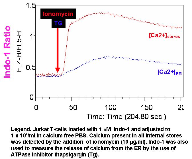
- Indo-1 detection of internal stores of calcium
- Effects of external calcium on Rhod-2 calcium flux
- Protocol