
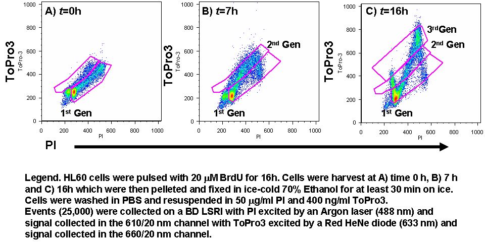
FRET can used to measure BrdU incorporation by the use of DNA dyes PI and ToPro3. The incorporation of BrdU is normally analysed in flow cytometry by labelling with a conjugate anti-BrdU mcab and PI to perform cell cycle analysis. This FRET based method relies upon the fact that when DNA is pulsed with BrdU the binding of PI and ToPro3 changes and are brought closer together (<10 nm) allowing FRET to occur resulting in an energy transfer from PI (donor) to ToPro3 (acceptor). Hence on a dot-plot of PI vs ToPro3, the BrdU containing cells have an enhanced ToPro3 signal compared to unpulsed cells. In longer time courses (>12h BrdU pulse) it is possible to track cell generations in a similar manner to that shown in CFSE proliferation assays, with advantage that cell cycle analysis data is collected.