
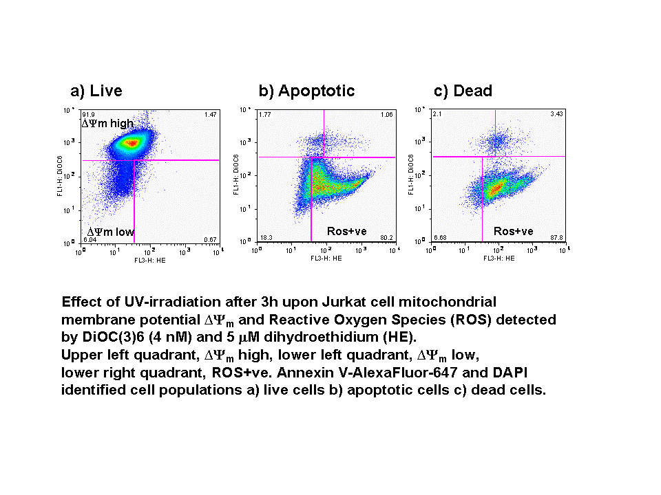
Organelle Function - Mitochondrial Membrane Potential (mmp)
The mmp is due a differential distribution of proteins on either side of the impermeable inner mitochondrial membrane. In apoptosis, irrespective of the stimulus, loss of mmp occurs with the formation of mitochondrial permeability transition pore (PT) or mitochondrial megachannel.
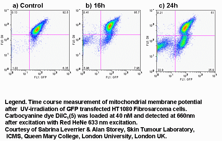
The fall in mmp can be measured flow cytometrically with a range of dyes all of which alter there fluorescence intensity with any change in mmp. These dyes include the carbocyanine molecules DiOC6(3) and DiIC1(5). Other cationic lipophilic dyes include CMXRos, TMRE and rhodamine 123.
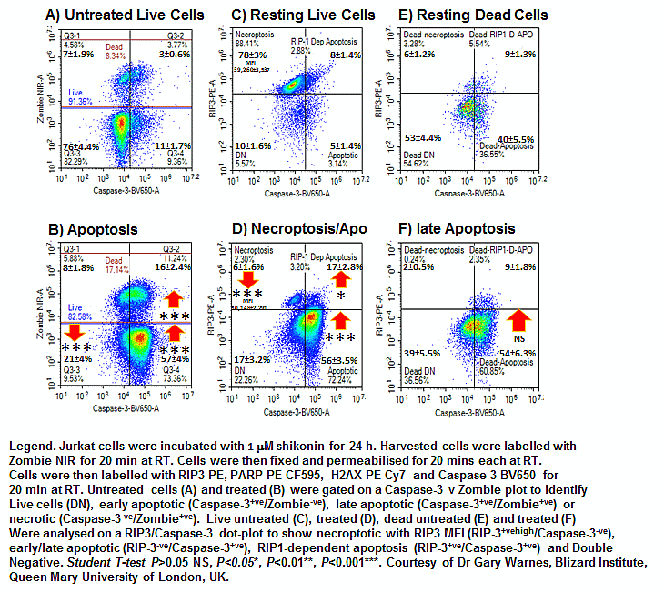
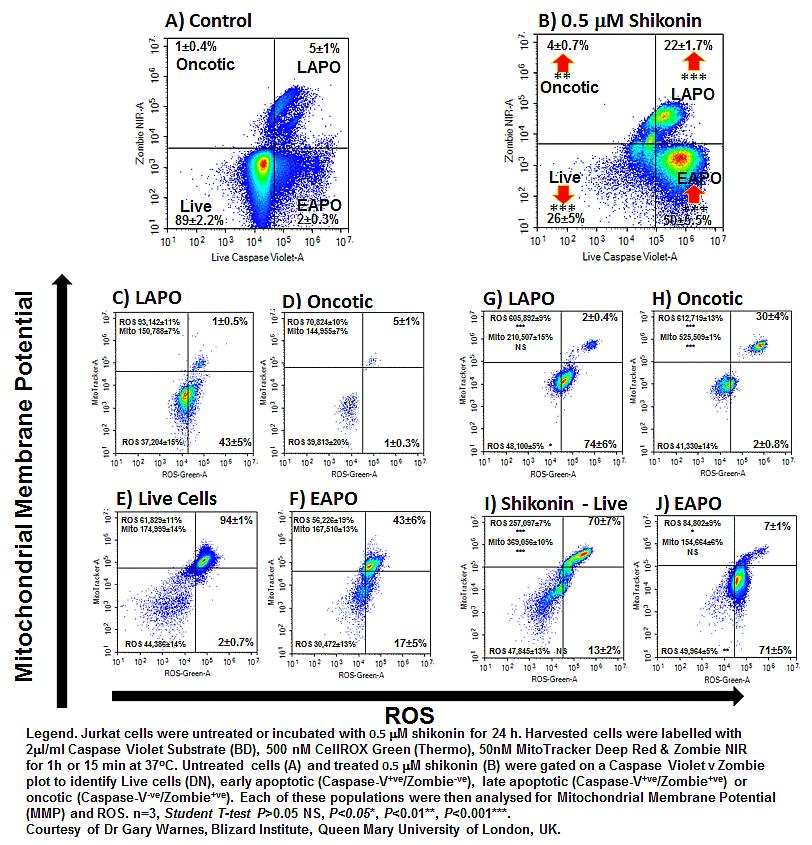
Shikonin induces necroptosis and apoptosis in Jurkat T cells which up-regulated RIP3 in live Caspase-3 negative cells. While early apoptotic cells not only only express activated caspase-3 but lack RIP3 expression, see figure. Shikonin also induces ROS in live cells which also show functioning mitochondria with hyper-polarized membrane potential (MMP), see figure. Early and late apoptotic cells show little MMP but increased ROS compared to untrated cells, see figure. Oncotic or cell viability+ve/Caspase Violet-ve cells after shikonin treatment show hyper-polarized MMP and a 5 fold increase in ROS, see figure.
Organelle Function - Reactive Oxygen Species
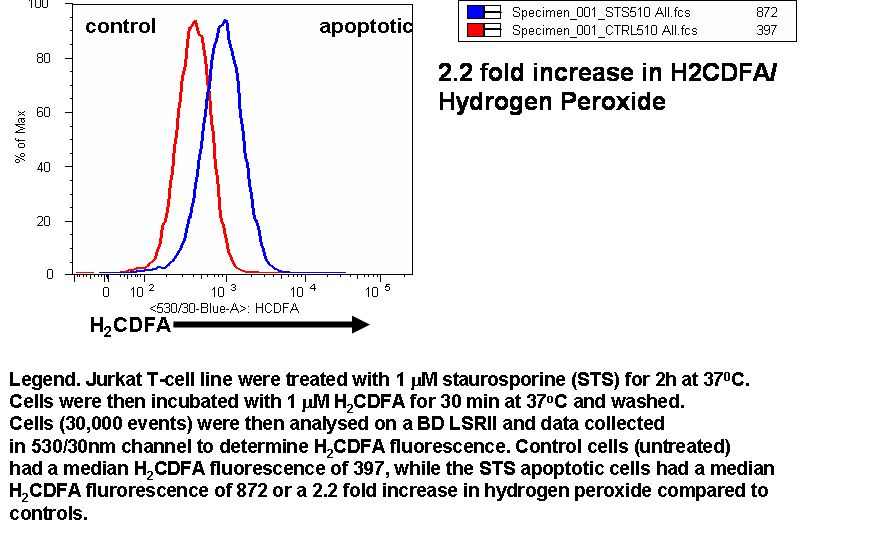
Reactive oxygen species include super-oxide, hydrogen peroxide and nitric oxide which are generated in small amounts during oxidative phosphorylation. Cells undergoing oxidative stress generate increasing amounts of superoxide and hydrogen peroxide.
There are numerous dyes available such as H2CFDA which are analogues of fluoroscein and dihydrogenated molecules of rhodamine 123 and Calcein AM that preferentially detect hydrogen peroxide.
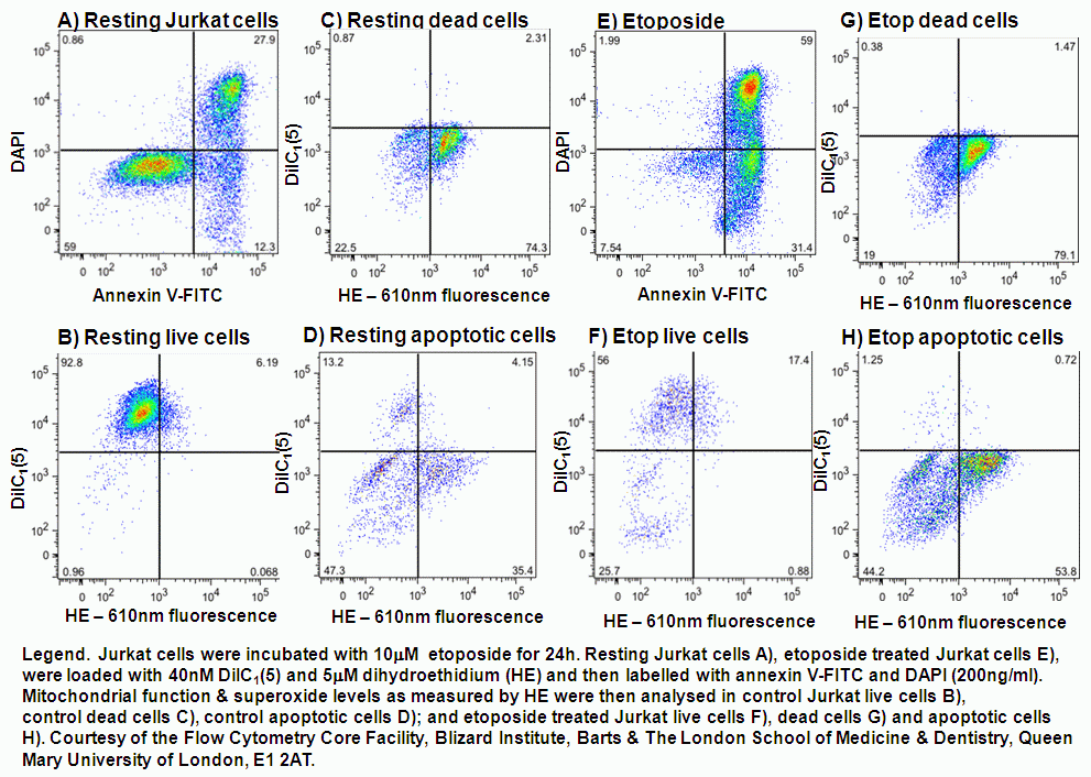
Superoxide is preferentially detected by dihydroethidium and Oxyburst Green H2CFDA.
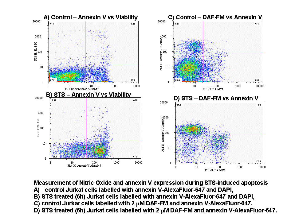
Nitric oxide is produced from L-Arginine by nitric oxide synthase. DAF-FM and DAF-FM acetate detect the presence of intracellular nitric oxide.