
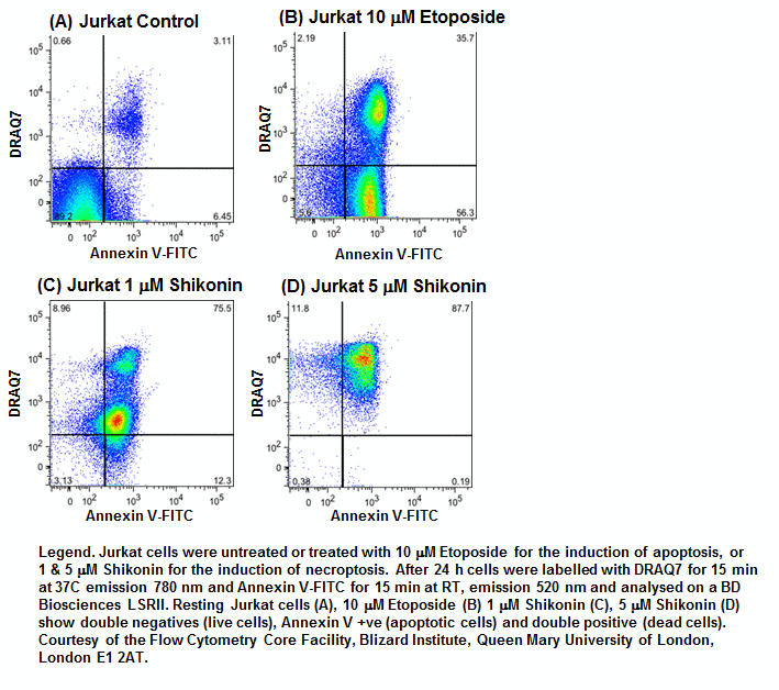
Classic apoptosis with activation of caspase-3 in the absence of RIP3 in cells without or with a permeable plasma membrane identifies early and late apoptosis respectively, see figure. Pre-labelling of cells with a fixable cell viability dye and subsequent fixing and permeabilisation allows intracellular labelling of active caspase-3 and RIP3 to identify these two stages of classic apoptosis. This is refinement of the now classic annexin V binding assay which although reasonably cheap and easy to use is not specific for any form of Regulated Cell Death (RCD).
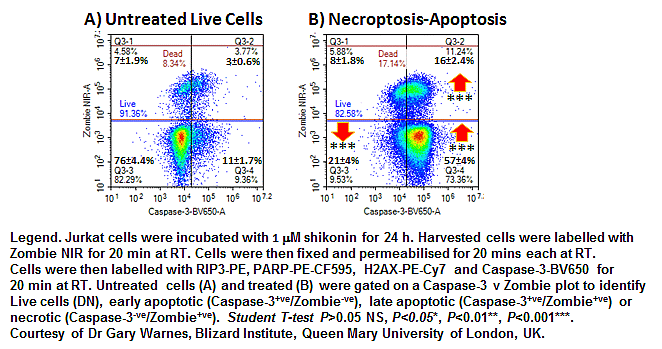
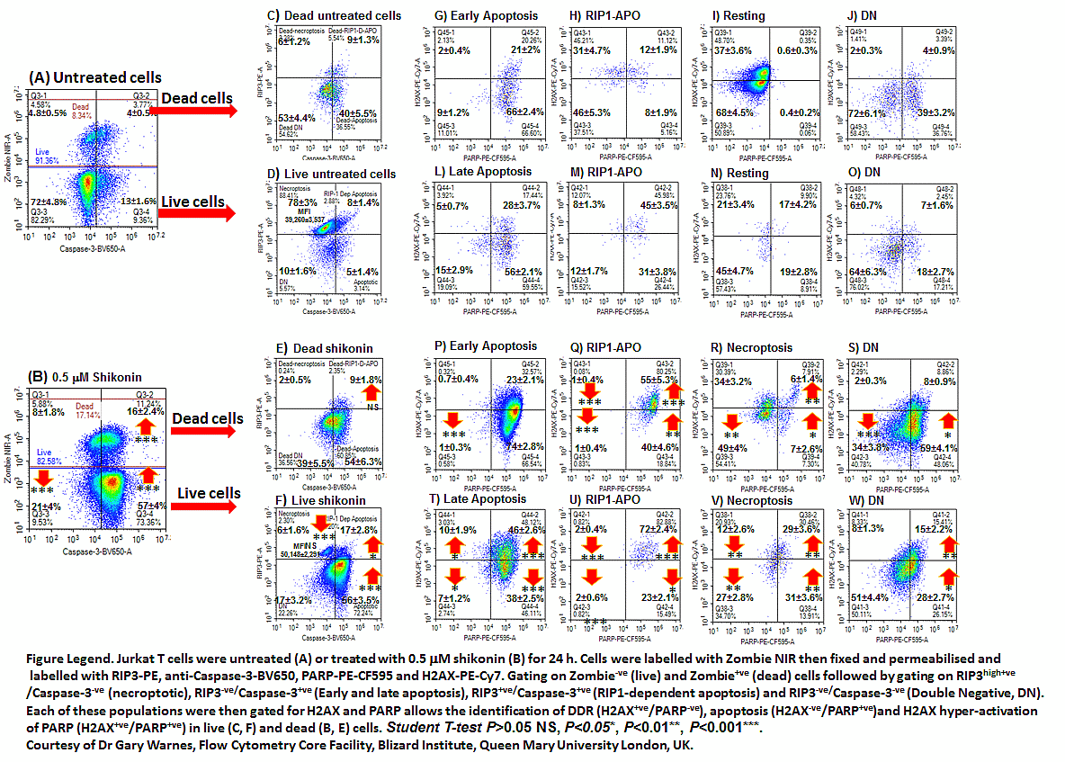
Cells during necroptosis auto-phosphorylate RIP1 which then phosphorylates and up-regulates RIP3 expression forming the necrosome. Mixed Lineage Kinase Like (MLKL) protein is then also phosphorylated which then forms pores within the cell plasma membrane resulting in cell necrosis. This up-regulation of RIP3 can be measured flow cytometrically with Caspase-3 being used to identify early and late apoptosis as well as RIP1-dependent apoptosis in both live and dead cells, see figure.
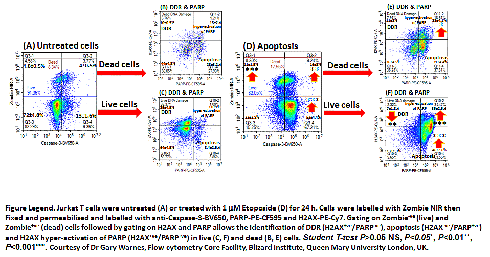
Etoposide induces apoptosis which can be immuno phenotyped using a fixable live-dead probe along with Caspase-3 and RIP3 as described above. Further immuno phenotyping with H2AX and cleaved PARP permits the identification of DNA Damage (H2AX+ve), apoptosis (PARP+ve), H2AX hyper-activation of PARP (H2AX+ve/ PARP+ve) and Double Negative (DN) cells, see figure.
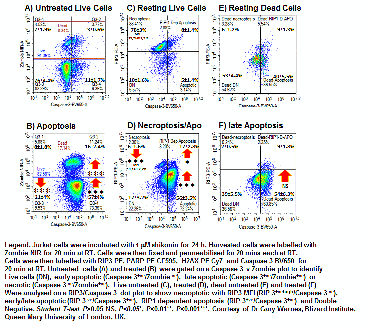
Jurkat cells treated with shikonin induces both necroptosis and classic caspase-3 dependent apoptosis producing populations with up-regulated RIP3/Caspase-3-ve (necroptotic), early and late apoptotic (RIP3-ve/Caspase-3+ve) , RIP1-dependent apoptosis in both live and dead populations. There are also cells that are double negative for both RIP3 and Caspase-3. All these populations have various levels of gamma H2AX+ve/PARP-ve (DNA Damage), hyper-activated PARP or parthanatos (H2AX+ve/PARP+ve), cleaved PARP (H2AX-ve/PARP+ve) or double negative, see figure.