Image - Barts and The London logo and link to home page Image - divider Image - divider
 
  Institute of Cell and Molecular Science
Flow Cytometry Core Facility equipment, images used with permission by BD
  Flow Cytometry Core Facility
  link Home link Instruments available link Flow cytometry link Uses of flow cytometry link Flow sorting link Links link Contacts
 

Microsomes


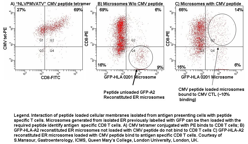
Isolation of GFP-HLA-A2 labelled Endoplasmic Reticulum (ER) can be sonicated into microsomes and identified by flow cytometry and loaded with CMV-peptide to identify antigen specific CD8-PE T-cells, see figure.

Publications.

DD Ateh, VH Leinster, SR Lambert, A Shah, A Khan, H Walklin, J Johnstone, N Ibrahim, M Kadam, Z Malik, M Girones, G Veldhuis, G Warnes, S Marino, I McNeish, JE Martins. The intracellular uptake of CD95 modified paclitaxel-loaded poly(lactic-co-glycolic acid) microparticles Biomaterials 32 (33), 8538-47, 2011.

 
Top


ER Microsomes

by Gary Warnes. © Queen Mary, University of London 2007
Institute of Cell and Molecular Science, The Blizard Building, Barts and The London School of Medicine and Dentistry, 4 Newark Street, London E1 2AT, UK Tel: +44 (0)20 7882 2483, Fax: +44 (0)20 7882 2200