To detect intracellular antigens it is necessary to permeabilise the cell. Many reagents are available commercially to facilitate this.
- Commercial permeabilisation reagents [PDF - 52KB]
There is a wide variety of different types of intracellular antigen that can be detected by flow cytometry, including:-
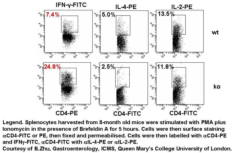
Lymphocytes and splenocytes involved in inflammatory or allergic responses produce a preponderance of IFNgamma or IL-4 respectively. KO murine splenocytes Th1Th2 responses were compared to wild type splenocytes. The KO showed an increased capacity to produce IFNgamma compared to the wild type response whilst IL-2 and IL-4 levels remained the same as the wild type.
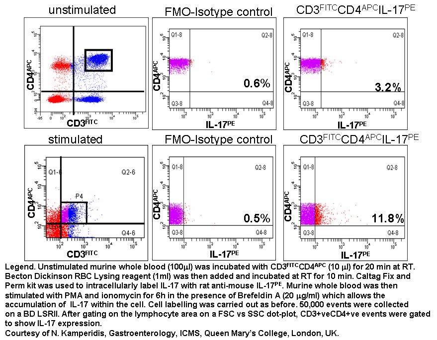
Detection of intracellular IL-17
Interleukins (e.g. IL1-17) can be measured intracellularly in lymphocytes after stimulation typically with PMA/ionomycin and blocking the Golgi apparatus with the antibiotic Brefeldin A or Monensin, see protocol.
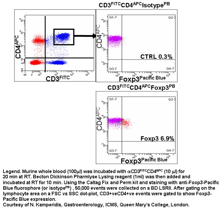
Foxp3 the self-reactive transcription factor, the master regulator involved in lymphocyte development and function of regulatory T cells can be measured intracellularly by flow cytometry, see protocol.
The intracellular detection of cyclins can be combined with cell cycle analysis, see cell cycle analysis section.
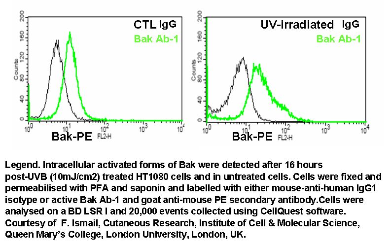
The pro-apoptotic factor, bak undergoes a conformational change when activated during apoptosis involving the mitochondria. The degree of bak activation can be determined intra- cellularly by use of mcab that detects the activated form of bak, see protocol.