
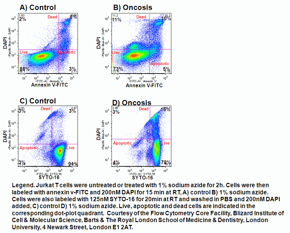
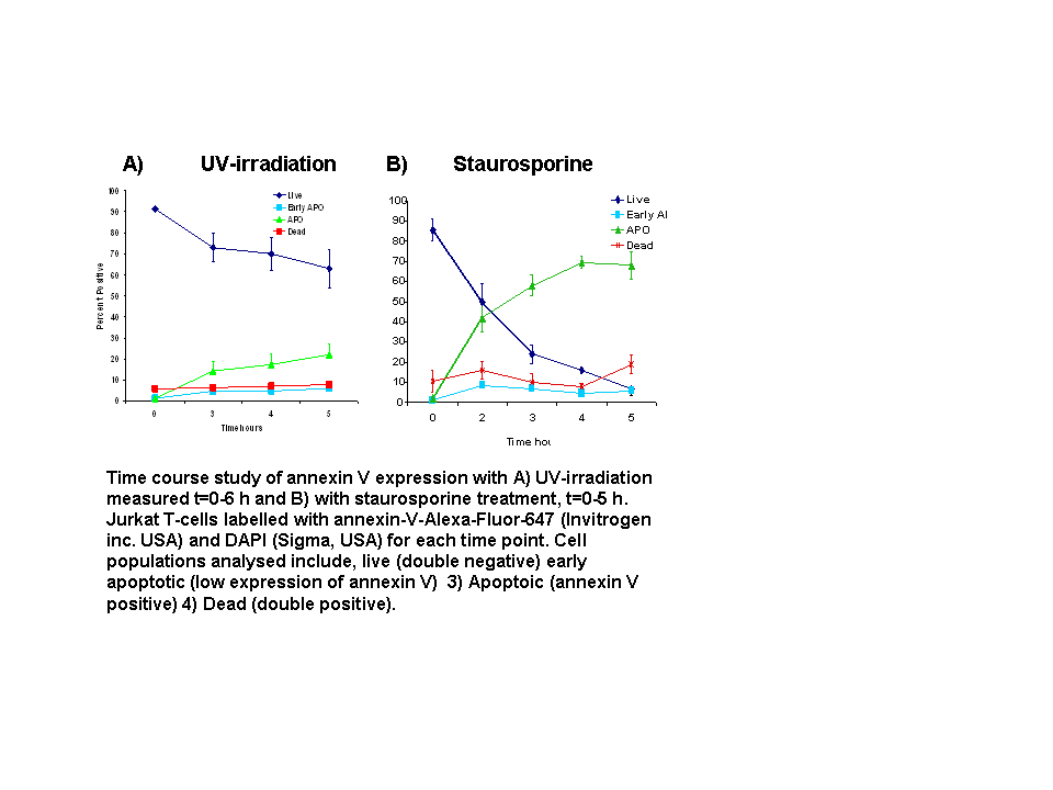
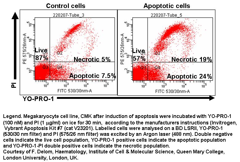
In normal cells, phosphatidylserine (PS) residues are found in the inner membrane of the cytoplasmic membrane. During apoptosis, the PS residues are translocated in the membrane and are externalized. Until recently oncosis has been studied by the use of annexin V binding to externalized phospha- tidylserine (PS) residues which is exposed in relatively few cells undergoing cell death by oncosis in comparison to that observed in cells undergoing apoptosis.
More recently the DNA binding dye SYTO 16 which has also shown to bind to the cytoplasm possible binding to mitochondria. In cells undergoing apoptosis the fluorescence intensity of SYTO 16 decreases before the plasma membrane loses its integrity making it a cheaper assay to detect apoptosis than annexin V when combined with cell viability dyes such as PI, DAPI, 7-AAD and DRAQ7. However recently it has been found that cells undergoing oncosis do not show this decrease in SYTO 16 before the cell membrane loses its integrity. Thus SYTO16 is a novel method for the detection of oncosis when combined with a viability dye such as DAPI, see figure.
- Model of plasma membrane
- Comparison of annexin V and SYTO 16 detection of oncosis
- Protocol-SYTO 16 assay
- Protocol-Annexin V assay