
Annexin-V - Cell membrane changes
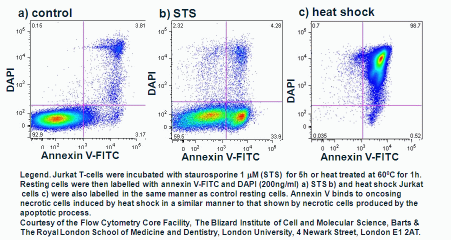
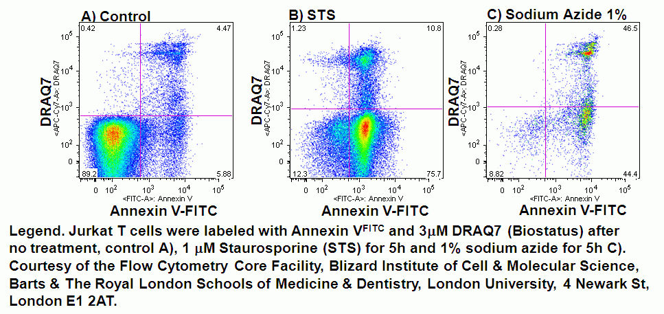
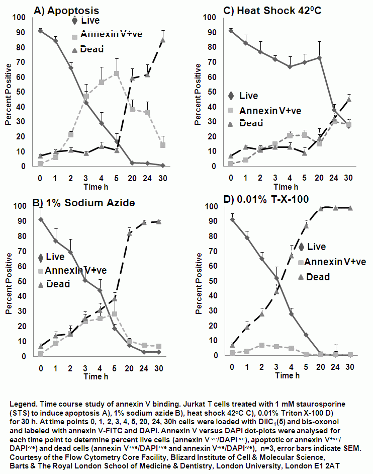
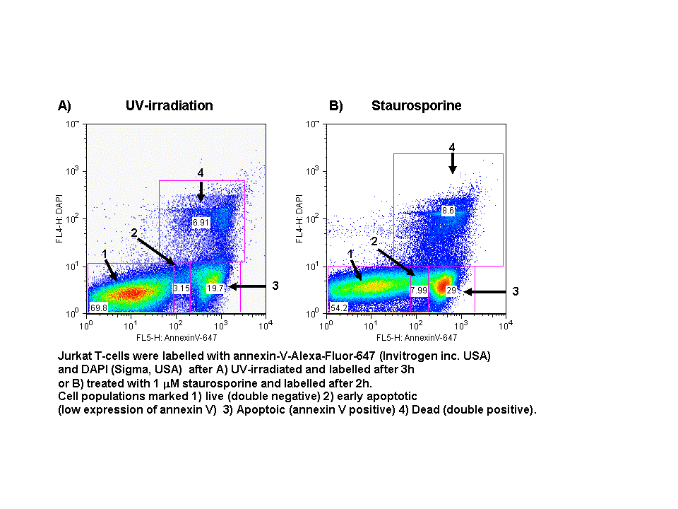
In normal cells, phosphatidylserine (PS) residues are found in the inner membrane of the cytoplasmic membrane. During oncosis, the PS residues are translocated in the membrane and are externalized. In general, this is not an early event in oncosis and the the role of PS externalization is unclear in oncosis. In oncosis, annexin V is also externalized during the early phase of oncosis; the cells then rapidly proceed to cell death and losing membrane integrity, see figure. Oncosis is induced by heat shock (56C or 42C), sodium azide (1% solution), Triton X-100 or drugs used to induce apoptosis if used at higher concentrations can induce oncosis. Annexin-V is a specific PS-binding protein that can be used to detect apoptotic cells. Annexin-V is available conjugated to a number of different fluorochromes. Annexin-V is a specific PS-binding protein that can be used to detect some oncotic cells. Annexin-V is available conjugated to a number of different fluorochromes including Alexa Fluor-647 and FITC. Numerous DNA binding viability dyes can be used in the annexin V assay including PI, DAPI and DRAQ7 (Biostatus).
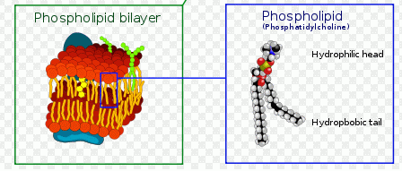
- Model of plasma membrane
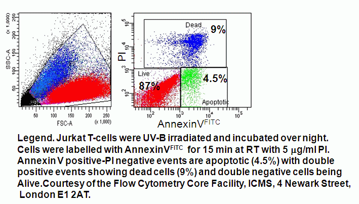
- Quadrant dot plot analysis of peripheral blood mononuclear cells with Annexin V-FITC and DAPI
- Annexin V Time Course Study
- Protocol