
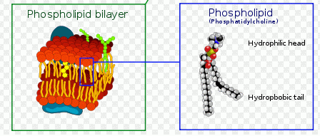
Annexin-V - Cell membrane changes
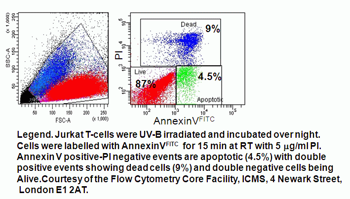
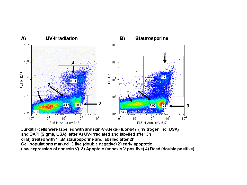
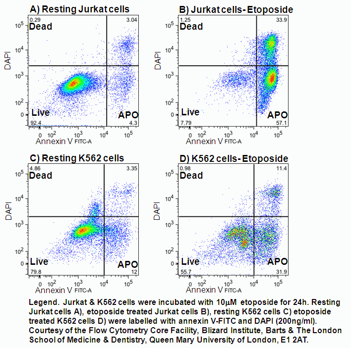
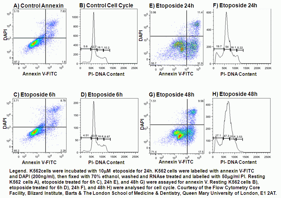
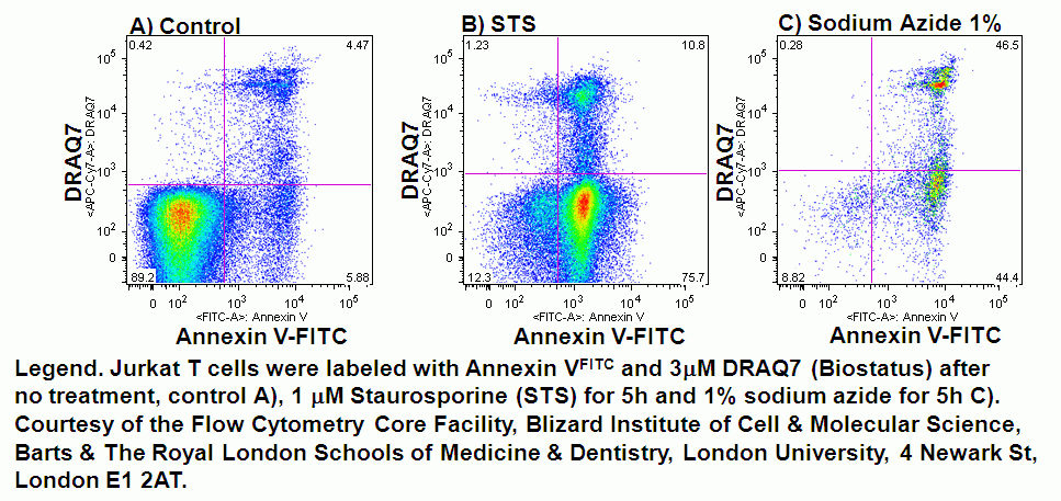
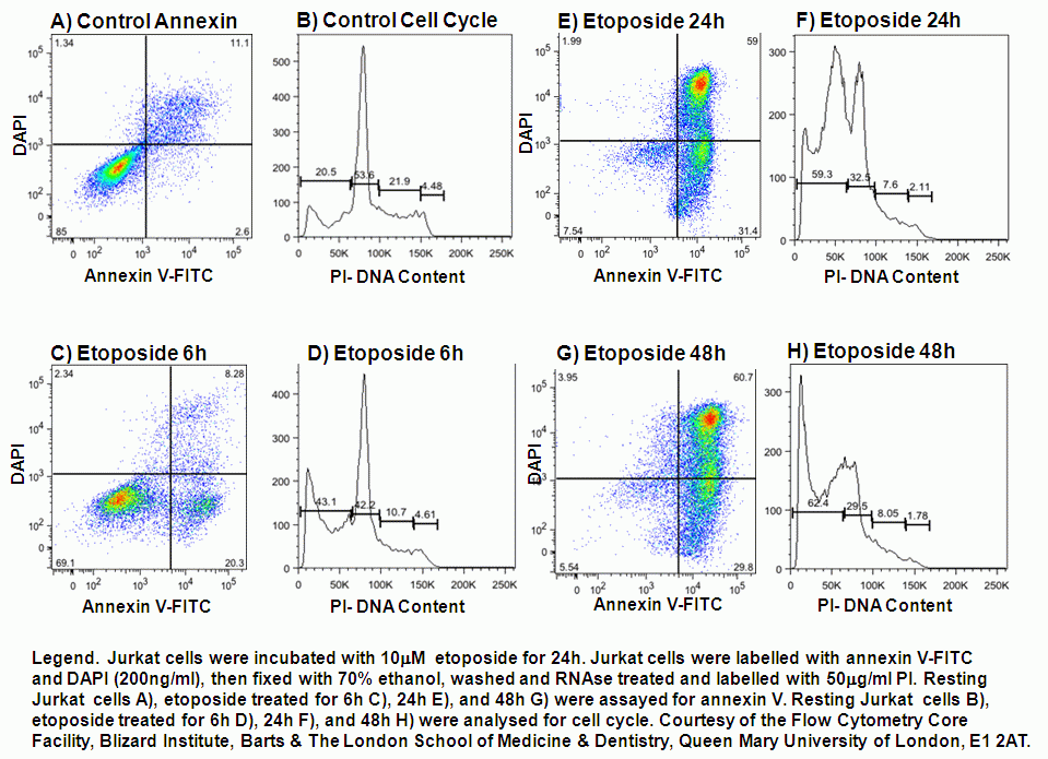
In normal cells, phosphatidylserine (PS) residues are found in the inner membrane of the cytoplasmic membrane. During apoptosis, the PS residues are translocated in the membrane and are externalised. In general, though not always, this is an early event in apoptosis and is thought to be a signal to neighbouring cells that a cell is ready to be phagocytosed. Annexin-V is a specific PS-binding protein that can be used to detect apoptotic cells. Apoptosis can be induced by UV-irradiation, as well as by numerous chemical treatments including the use of pan-kinase c inhibitor, staurosporine at high concentration and etoposide phosphate, a topoisomerase II inhibitor which stops the action of DNA unwinding enzyme topoisomerase II during DNA synthesis causing DNA breaks to occur during S phase resulting in apoptosis which can be observed with Jurkat and K562 cells, that have been previously analysed for annexin V and cell viability.
Annexin-V is available conjugated to a number of different fluorochromes including Alexa Fluor-647 and FITC. Numerous DNA binding viability dyes can be used in the annexin V assay including PI, DAPI and DRAQ7 (Biostatus). The Millipore Muse mini flow cytometer can be used to determine apoptosis levels in a cell sample by labelling with annexin V-PE and 7-AAD as the viability dye, see figure.
- Model of plasma membrane
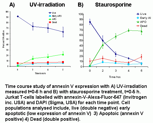
- Apoptosis induction by UV-irradiation
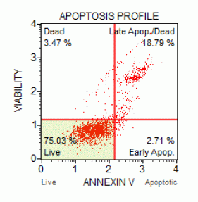
- Quadrant dot plot analysis of Jurkat T-cell line with Annexin V-Alexa-Fluor-647 and DAPI
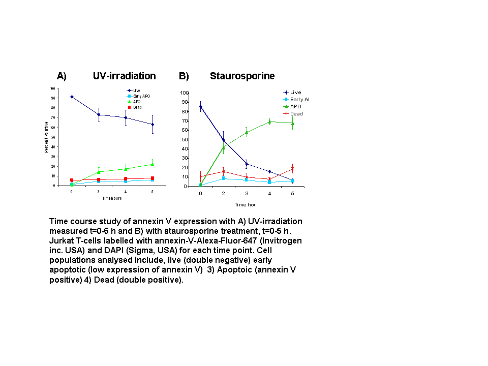
- Annexin V Time Course Study
- Protocol
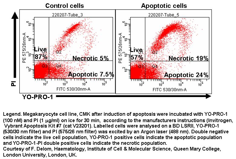
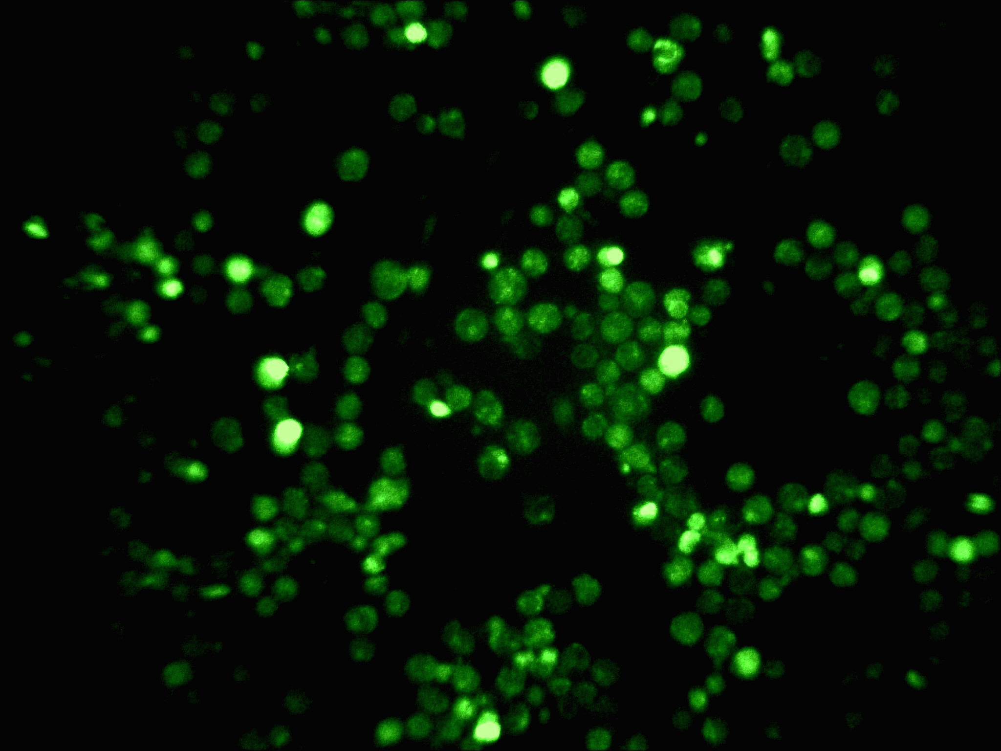
Instead of PI, TO-PRO-3 can be used to specifically identify the dead cells. Also YO-PRO-1 (Invitrogen) a fluorochrome that emits in the green region, can be combined with propidium iodide to identify dead cells. Cells in early apoptosis are unable to pump out YO-PRO-1 but are still not permeable to other dead cells discriminatory dyes. Live cells are not fluorescent or double negative, apoptotic cells are green (YO-PRO-1+ve) and dead cells are double positive PI+ve-YO-PRO-1+ve.