
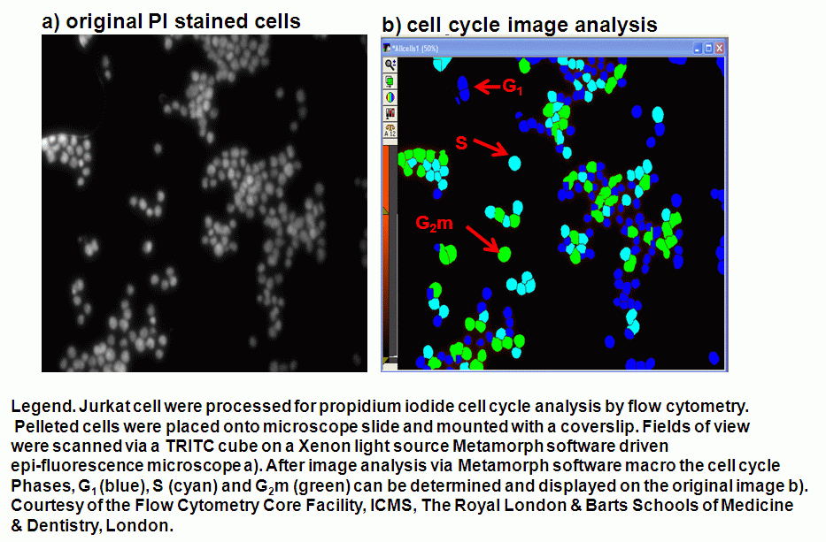
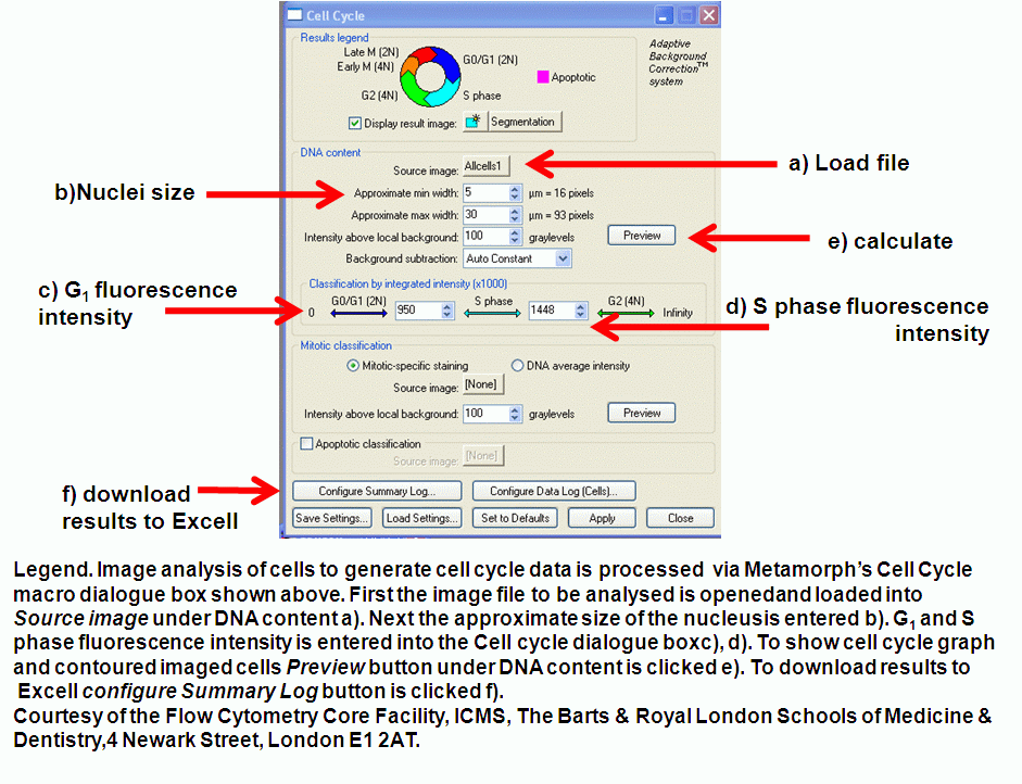
The determination of the cell cycle analysis is a well known technique in flow cytometry. The Core Facilities at ICMS has a Metamorph epi-fluorescent deconvolution system with a software macro to determine the cell cycle stage of cells on slides or in culture.
Jurkat cells stained with DNA binding dyes such as propidium iodide (PI) or DAPI can be analysed by Metamorph to determine the percentage of cells in G1, S, G2m and apoptosis.
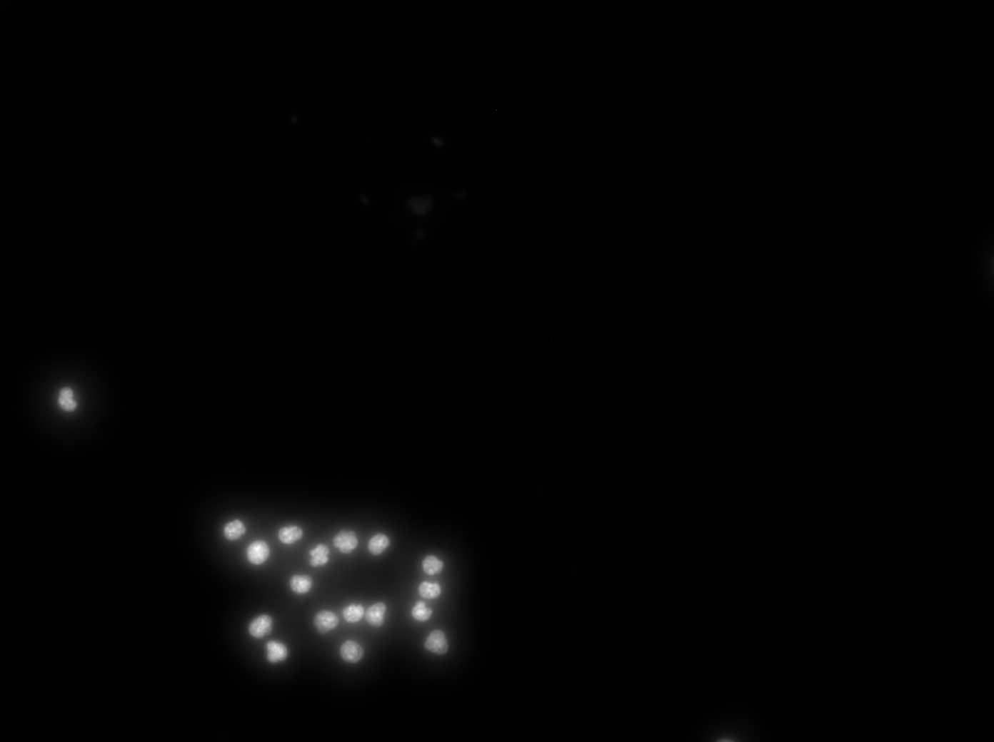
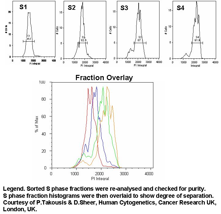
Jurkat cells were sorted on a BD FACSAria into G1, S phase and G2m cells. Centrifuged cells were placed onto microscope slides and imaged at the same exposure and magnification (x20). This allows the system to be calibrated so that unsorted cells can be analysed using the Metamorph cell cycle analysis macro.