Live Go cell cycle analysis with Hoechst 33342 and Pyronin Y
Flow cytometry may be used to determine the level of quiescence or senescence in a cell population. The biological processes involved in quiescence and senescence are different in that senescence has to be induced by g-irradiation or cyclin-dependent kinase inhibitor, CDKNA1A and 2A putting cells into Go phase of the cell cycle. This cell cycle arrest after several days is irreversible and the cells grow in size without cell division and acquire SA-B-Gal staining and after a profound reorganization of chromatin structure and the endomembrane system enter a secretory state, releasing a mass of factors to the external environment called the senescence-messaging secretome (SMS) or senescence-associated secretory phenotype (SASP).
Cell quiescence as opposed to senescence is typically induced by serum starvation and cells enter the Go phase of the cell cycle but the process is reversible, the cells entering G1 when nutrients are supplied. Thus the of RNA binding dye, Pyronin Y to determine Go phase cells is probably more useful in detecting quiescent cells as opposed to senescence where the cells are actively producing protein from mRNA. Consequently senescent cells will contain more RNA than quiescent cells.
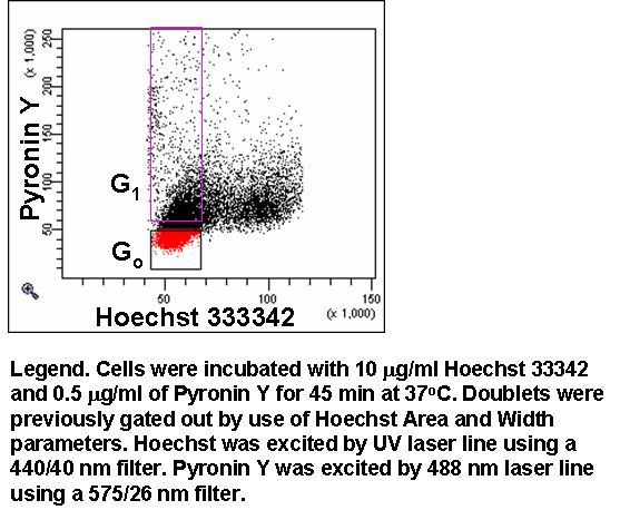
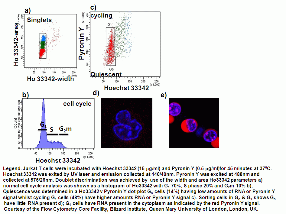
Go and G1 can be separated by use of Pyronin Y (Sigma) which preferentially binds to RNA and Hoechst 33342 (Sigma) which binds to A-T base pairs. Cells were loaded with Hoechst 33342 at 10 mg/ml and Pyronin Y at 0.5 mg/ml by incubating for 45 mins at 37C. Pyronin Y is excited at 488 nm and emits at 575 nm, while Hoechst 33342 is excited UV lasers (350-360 nm), near UV line (375 nm) or violet diode (405 nm).
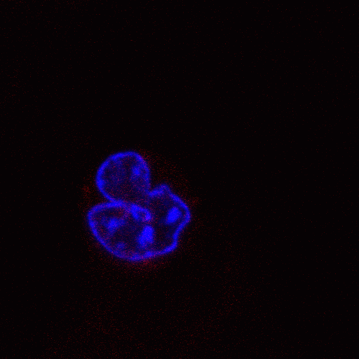
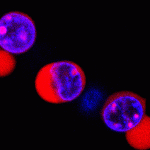
Examples of fibroblasts, K562 cells, Jurkat T cells stained with Hoechst 33342 and Pyronin Y show the presence of Go cells with little or no RNA present whereas the G1 cells show high level expression of RNA. The small amount of RNA present in Go cells was so low using the same PMT settings on a Zeiss confocal no RNA was visible. Therefore the Go cell imaging was carried out at 250 volts higher than the G1 cycling cells. The confocal imaging of Pyronin Y in Go quiescent and G1 cycling cells clearly shows a large difference RNA content of these cells in the two stages of the cell cycle.
Fixed cell analysis
The cell cycle protein associated with senscence is p16 and this can be analysed flow cytometrically against cell cycle, see figure