
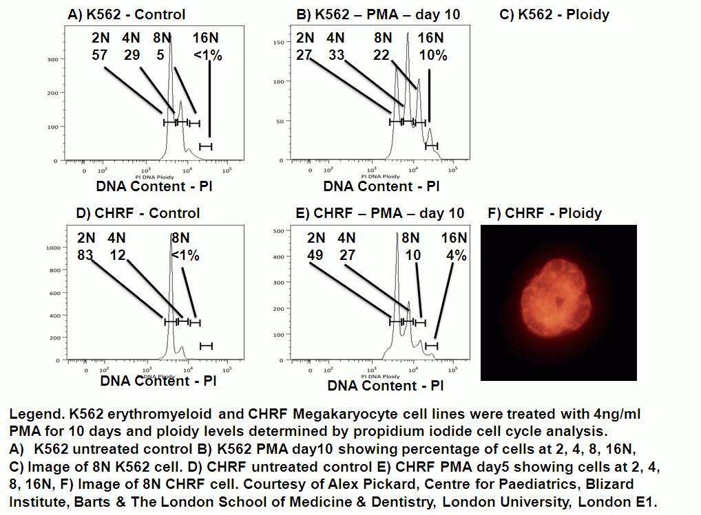
Ploidy or endoduplication occurs when cells in G2M do not separate after DNA synthesis and proceed to synthesis DNA again become 8N cells. This can be observed in cell lines quite often with a small population of these going onto synthesis DNA again becoming 16N.
Classically Megakarocytes were analysed in this way, as these platelet progenitor cells can be as large as 256N with peaks at 128N, 64N, 32N, 16N and 8N. The mature very large Megakaryocyte then releases up to 3,000 platelets. Log scale fluorescence parameters are used to detect cells greater than 16N on modern digital instrumentation.
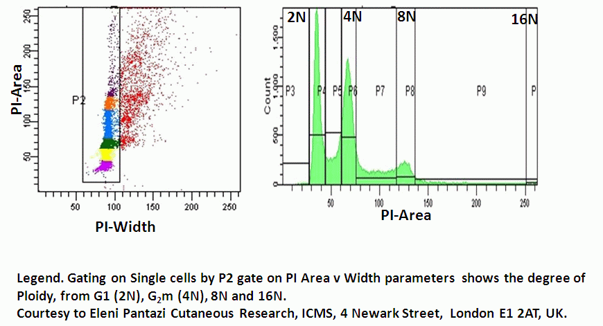
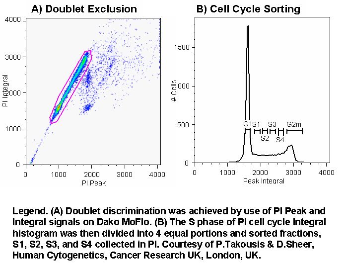
NTERT cells were analysed for ploidy by the standard cell cycle protocol by fixing cells in 70% ice-cold ethanol, washing and incubating with RNAse and propidium iodide (PI). The linear voltage on the 610/20nm channel was adjust so a doublet discrimination gate was placed on the Area vs Width parameter placing G1 at channel 25,000 on the BD FACSDiva software. This allows 2N, 4N, 8N and 16N to appear on a linear scale. For cells with ploidy greater than 16N log scaling should be used.
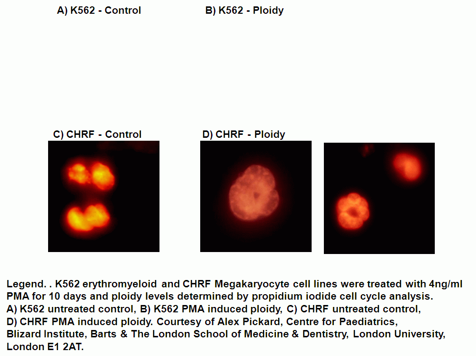
K562 erythromyeloid cell line and CHRF cells can be induced to become polyploid after PMA treatment. The degree of ploidy can be determined by fixing cells in 70% ice-cold ethanol for at least 1 hour, washing in PBS twice and treating cells with RNAse and staining with 50ug/ml PI, see figure. Epi-fluorescence imaging with an 100x objective shows lobed nuclei after PI treatment, see figure.
Publications
E Pantazi, E Gemenetzidis, G Trigiante, G Warnes, L Shan, X Mao, M Ikram, MT Teh, YJ Lu, M Philpott.GLI2 induces genomic instability in human keratinocytes by inhibiting apoptosis. Cell Death Dis. Jan 30.5:e1028 doi 10.1038/cddis 2013.535 2014.