
Although propidium iodide is probably the most commonly used dye to quantitatively assess DNA content, there are several different dyes that are available which bind stoichiometrically to DNA. These may be divided as follows:
- UV excited: Hoechst 33342, Hoechst 33258, DAPI
- 457nm excited: Mithramycin
- 488nm excited: Propidium Iodide, 7-Aminoactinomycin
- 532nm excited:Propidium Iodide
- D, SYTOX Green, DRAQ5
- 633nm excited: TO-PRO-3 Iodide
- Violet (405nm) excitation of Hoechst 33342
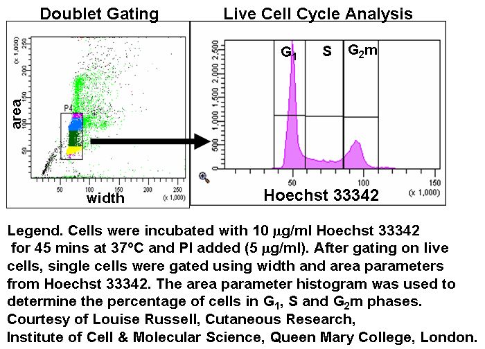
One important aspect of DNA analysis is the ability of the cytometer to exclude cell doublets. In a cytometer where the beam diameter is larger than the nuclear size, we can look at pulse width versus pulse area to exclude cell doublets - a true G2 cell will have a smaller width than two G1 cells passing through the beam together consecutively.
This technique is based on the premise that cells in G0 or G1 phases of the cell cycle possess a normal diploid chromosomal, and hence DNA content (2n) whereas cells in G2 and just prior to mitosis (M) contain exactly twice this amount (4n). As DNA is synthesized during S-phase, cells are found with a DNA content ranging between 2n and 4n. A histogram plot of DNA content against cell numbers gives the classical DNA profile for a proliferating cell culture.
- Protocol - PI staining
- Use of RNase with PI protocol
- Protocol - DAPI staining
- Protocol - 7-AAD staining
UV excitable DNA dyes such as DAPI and Hoechst 33342 are normally excited with a UV laser (350-360nm) for cell cycle analysis. There are available commercial dyes for excitation with a violet 405nm laser diode, however if cells are fixed with 4% fresh paraformaldehyde solution (Polysciences) for 1 hour then labeled with 10 ug/ml Ho33342 a good cell cycle profile can be obtained, see figure. This allows the user to analyze cells for GFP content versus cell cycle without loosing significant amounts of GFP or use other channels for antibody fluorochrome detection with cell cycle analysis without the need for a UV laser line.
Triton X-100 (final 0.25% solution v/v) can be used to permeabilse cells within 5 minutes. DAPI (1ug/ml), Hoechst 33342 (10 ug/ml), PI (50 ug/ml) or 7-AAD (25 ug/ml) added and cells analysed for cell cycle analysis. DAPI and Hoechst 33342 can also be used to determine the cell cycle of cells with a violet 405nm laser diode giving CVs of the G1 peak of 6% as opposed to <5% when using a UV laser, see figure. The use of PI has the disadvantage in that RNASe cannot be used giving a falsely high S phase determination, see figure.