
Organelle Function - Reactive Oxygen Species
Reactive oxygen species (ROS) include a variety of molecules not only superoxide anion and hydrogen peroxide but singlet oxygen, peroxynitrite anion, hydroxyl radical and nitric oxide. These ROS molecules are produced normally and in disease states.
Superoxide is produced during phagocytosis and can be detected by OxyBURST Green H2DCFDA or Green H2HFF BSA. These dyes operate in a similar manner to the hydrogen peroxide sensitive dyes Carboxy-H2DCFDA and CMH2DCFDA by increasing in fluorescence in the presence of superoxide.
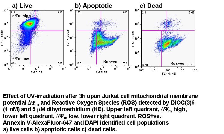
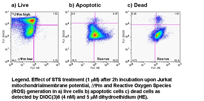
Superoxide is also produced during apoptosis and can be detected by dihydrogenated version of ethidium, dihydroethidium. This dye is oxidized by superoxide releasing ethidium which then fluoresces when bound to cellular DNA, see figure. Etoposide is shown to produce more superoxide in K562 cells than Jurkat T-cells after 24h of treatment, see figure.
Similarly dihydrorhodmaine 123 is oxidized preferentially by hydrogen peroxide the free rhodamine 123 then fluoresces upon binding to mitochondrial membranes.
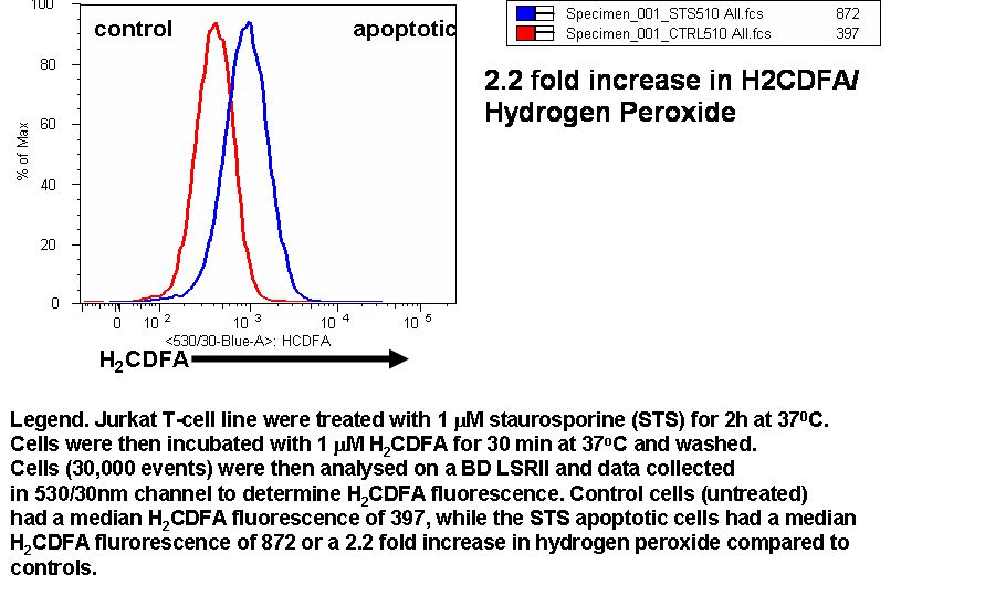
Hydrogen peroxide as mentioned above can be detected by H2CFDA and CMH2DCFDA which are acted upon by intracellular esterases the free fluoroscein molecules then interact with hydrogen peroxide generating an increase in fluoroscein signal.
The effect of oxygen levels with or without drug treatment on cell ROS levels as detected by CDFA was compared using the rectal carcinoma cell line, C80. Cells showed great differences generation of ROS when treated with drugs under normoxic or hypoxic conditions, see figure.
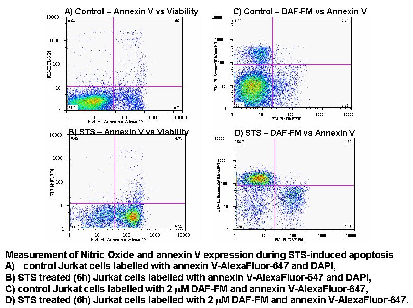
Nitric Oxide produced during apoptosis from L-Arginine by the action of nitric oxide synthase can be detected by loading cells with fluoroscein analog DAF-FM which increases in fluorescence upon binding nitric oxide.
Protocols:- Superoxide HE; Nitric Oxide DAF-FM, Hydrogen Peroxide H2CDFA