
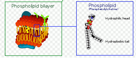
Plasma membrane changes
In normal cells, phosphatidylserine (PS) and phosphatidyl-ethanolamine (PE) residues are found in the inner membrane of the cytoplasmic membrane. During apoptosis, the PS and PE residues are translocated in the membrane and are externalised resulting in a change in surface charge of the outer leaflet of the plasma membrane. In general, though not always, this is an intermediate event in apoptosis and is thought to be a signal to neighbouring cells that a cell is ready to be phagocytosed. Annexin-V is a specific PS-binding protein that can be used to detect apoptotic cells by binding to PS. In general, this is not an early event in autophagy and the the role of PS externalisation is unclear in autophagy. Interestingly the phospholipid, phosphatidyl- ethanolamine (PE) plays a crucial role in the formation of the autophagosome during the induction phase of the autophagic response in that it lipidates the microtubule LC3 in the double membrane of the autophagosome to form LC3B which is used as a marker for autophagosomes and hence the detection of autophagic cells.
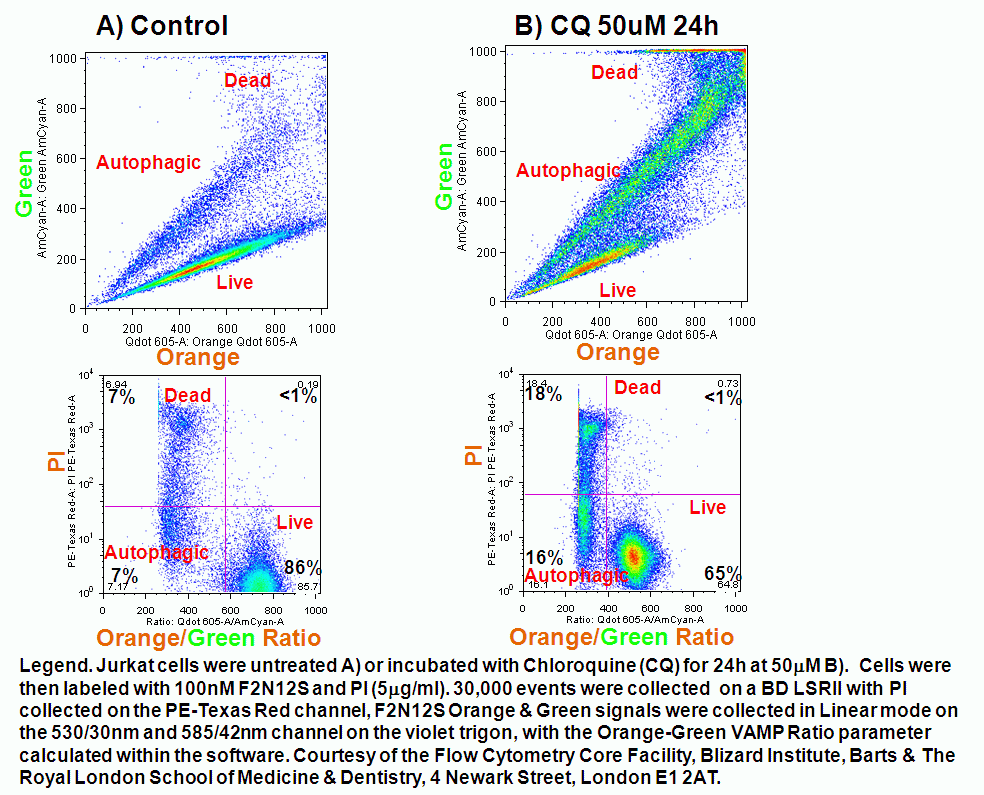
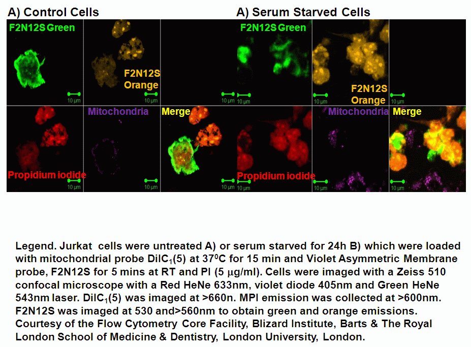
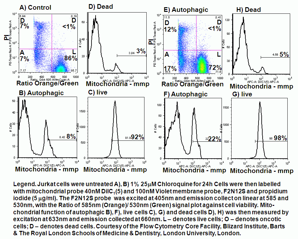
The autophagic inducing agent, chloroquine also induces apoptosis and thus PS is externalised to the outer leaflet of the plasma membrane and thus the annexin V assay or Invitrogens Violet Ratiometric Asymmetry Probe can be used to detect these cells undergoing apoptosis during the autophagic response to chloroquine which ultimately blocks the autophagic response by it's chemical characteristic of being a weak base which inhibits the fusion of lysosomes to the autophagosome and thus inhibits the completion of the autophagic process. Serum starvation can also induce autophagy in cells and the violet ratiometric asymmetry probe was imaged by confocal microscopy to show this type of autophagic process, see figure.
Violet ratiometric asymmetry probe
The voltage change associated with the externalisation of PS and PE can be detected by plasma membrane probe Bis-oxonol DiBAC4(5) and a new violet ratiometric membrane asymmetry probe, 4'-N,N-diethylamino-6-(N,N,N-dodecyl-methylamino-sulfopropyl)-methyl-3-hydroxyflavone or F2N12S is sensitive to changes in the potential difference or charge on the outer surface of the plasma membrane. The dye has an excited-state of intramolecular proton transfer or ESIPT reaction and in response to changes in the surface charge of the plasma membrane of cells emits fluorescence at two emissions of 530 and 585nm or green and orange fluorescence after excitation with a violet 405nm laser diode. Ratiometric analysis of the 2 emissions with the PMTs switched to linear gives all the advantages of ratiomtric probes over single colour probes in that detected changes are independent of probe concentration, cell size and instrument variation in laser outputs and sensitivity of PMT detectors.
In chloroquine induced autophagy the surface charge of the outer plasma membrane changes which F2N12S can measure by alterations in the intensity of the two emissions, which emit more at 530nm (green) and less at 585nm (orange). Thus ratioing the 585nm emission by the 530nm emission electronically and plotting this Ratio parameter against cell viability shows the presence of apoptotic cells by a reduced Ratio signal. Dead cells also have a reduced Ratio signal compared to healthy live cells, see figure.
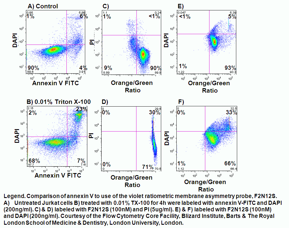
The F2N12S violet ratiometric membrane asymmetry probe is loaded into cells at RT for 5 minutes at 100nM concentration. Cells are not washed after this reaction but must be read within 30 minutes of loading cells. Then annexin V needs to be labeled before F2N12S, if a comparison of the two methods is required. Also although the emission spectra from F2N12S is at 530 and 575nm after 405nm excitation, DAPI with an emission spectra of 461nm cannot be used as a cell viability marker as there appears to be quenching of signals, see figure.
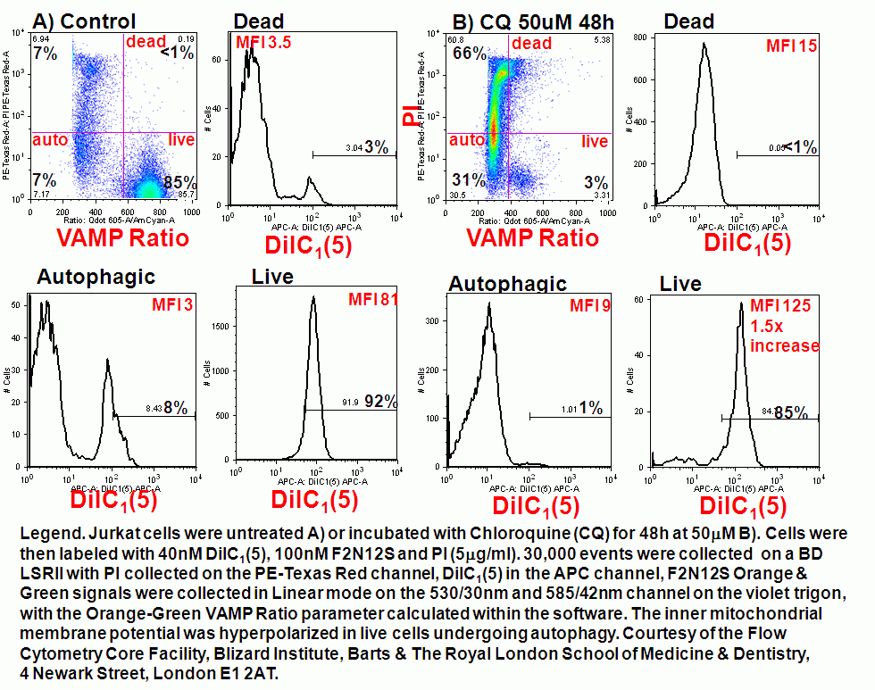
In addition other biological events can also be measured simultaneously such as mitochondrial membrane potential during autophagy.
- Model of plasma membrane
- Violet membrane asymmetry probe ratio
- Violet membrane asymmetry probe and mitochondrial function
- Protocol for F2N12S probe