
Role of electron microscopy in the characterization of autophagy
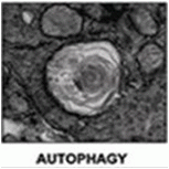
Autophagy was originally characterized in 1963 by De Duve with electron microscopy studies showing the presence of double membrane bound structures termed auto-phagosomes and single membrane vesicles terming autolysosomes or autophagolysosomes, see figure.
Light microscopy imaging of autophagic granules
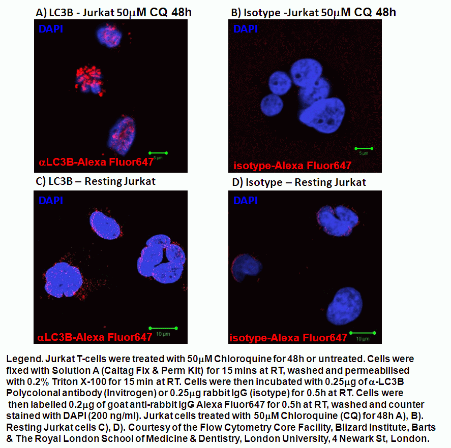
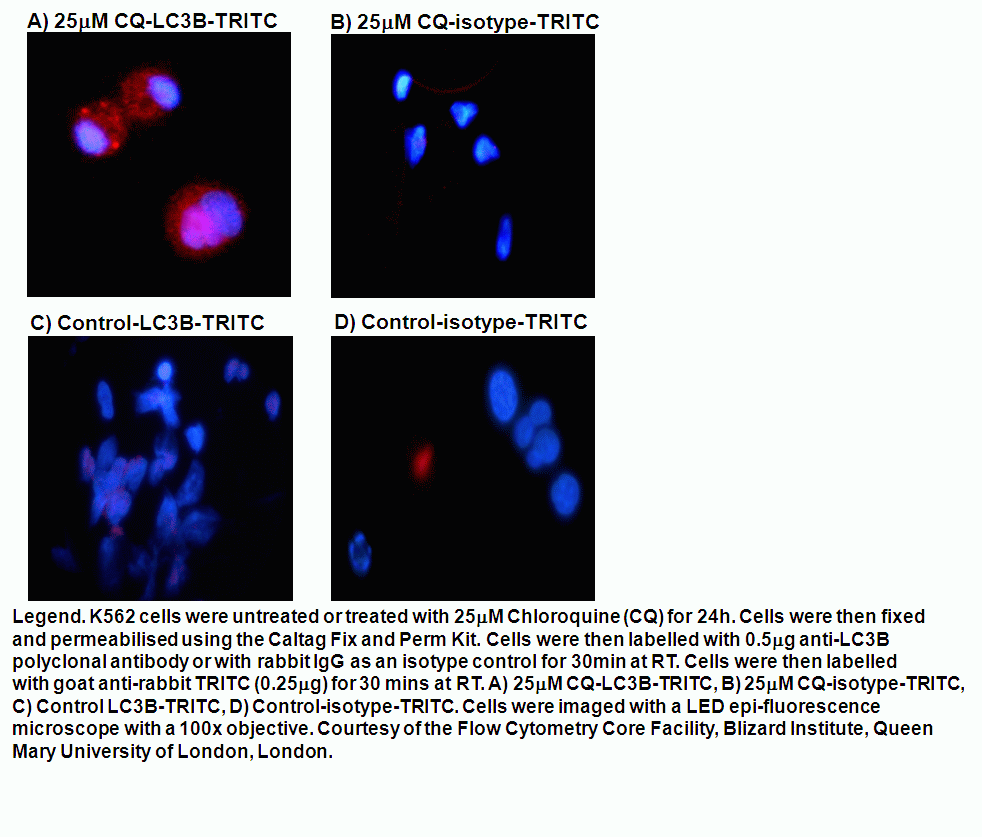
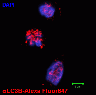
IThe current methods of determining the presence of autophagy or cells undegoing autophagic flux is based on the fact the microtubule protein, LC3 normally present in the cytoplasm, but during the autophagic process is cleaved and lipidated with the phospholipid, phosphatidylethanolamine (PE) to form LC3B in autophagosomes, see figure. After fusion of the autophagosome with lysosomes, to form the single membrane bound, autolysosome the the internal LC3B is reduced. The LC3B can be tagged with GFP (fluorescence is acid sensitive) and RFP (acid resistant) and imaged by fluorescence microscopy or analyzed flow cytometrically. The LC3B levels can also be measured by the use of polyclonal LC3B antibody tagged in this case with Alexa Fluor 647, which can then be analysed flow cytometrically. Both these methods can be analyzed flow cytometrically removing the requirement for time consuming image analysis of the number and level of LC3B molecular expression, see autophagosome section.
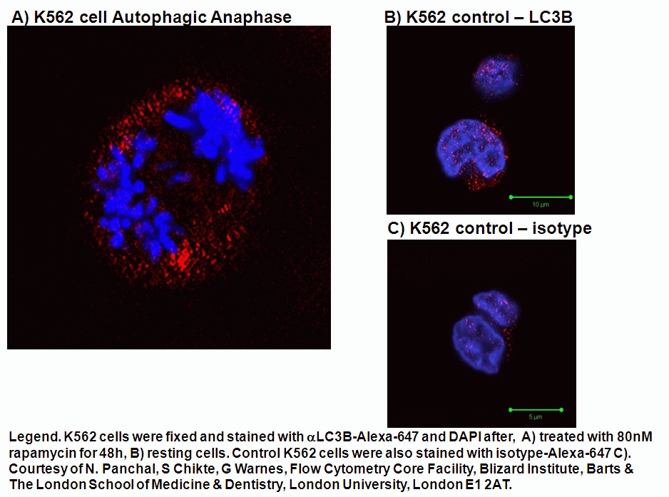
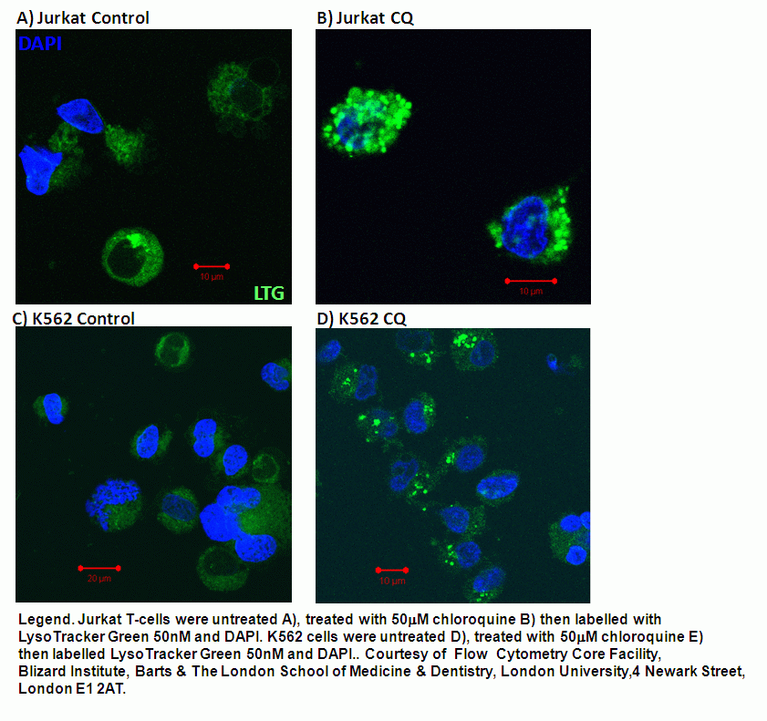
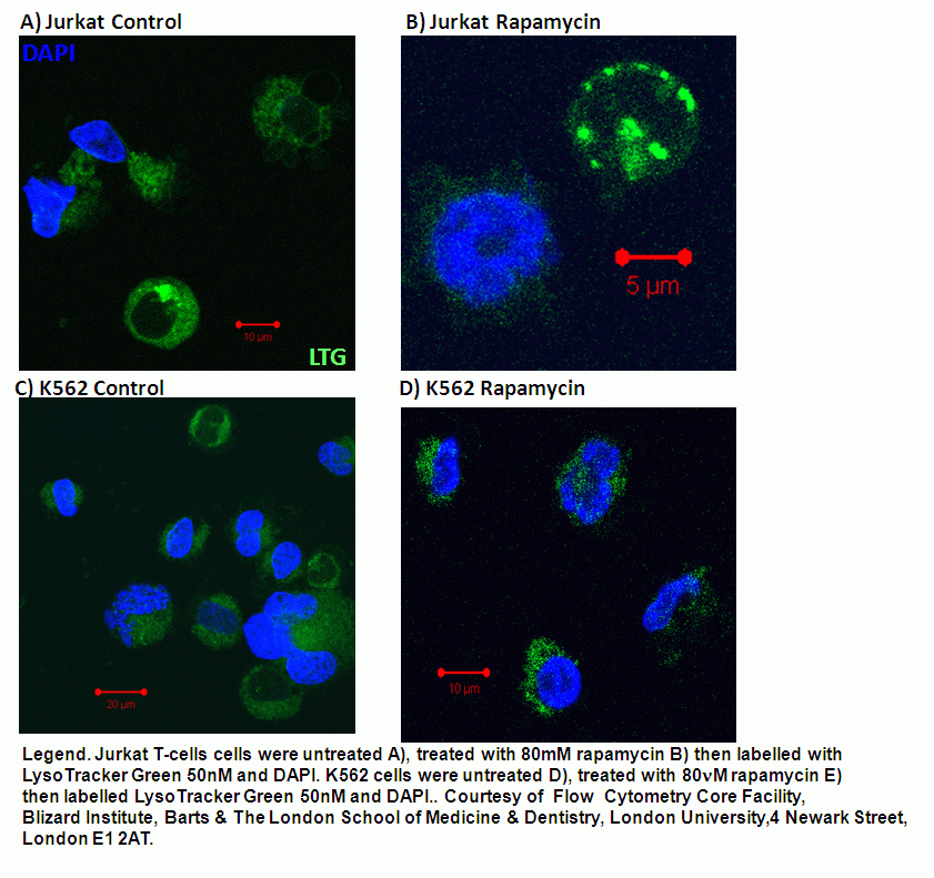
Interestingly chloroquine by it multiple mode of action results in the swelling of autophagosomes, see figure. Whilst rapamycin induction of autophagy results in smaller autophagosomes being detected by light microscopy, see figure.
LysoTracker dyes such as the Invitrogen LysoTracker Green and Red whose intensity have also been reported to increase during autophagic flux, see figure.
CMA - Chaperone Mediated Autophagy
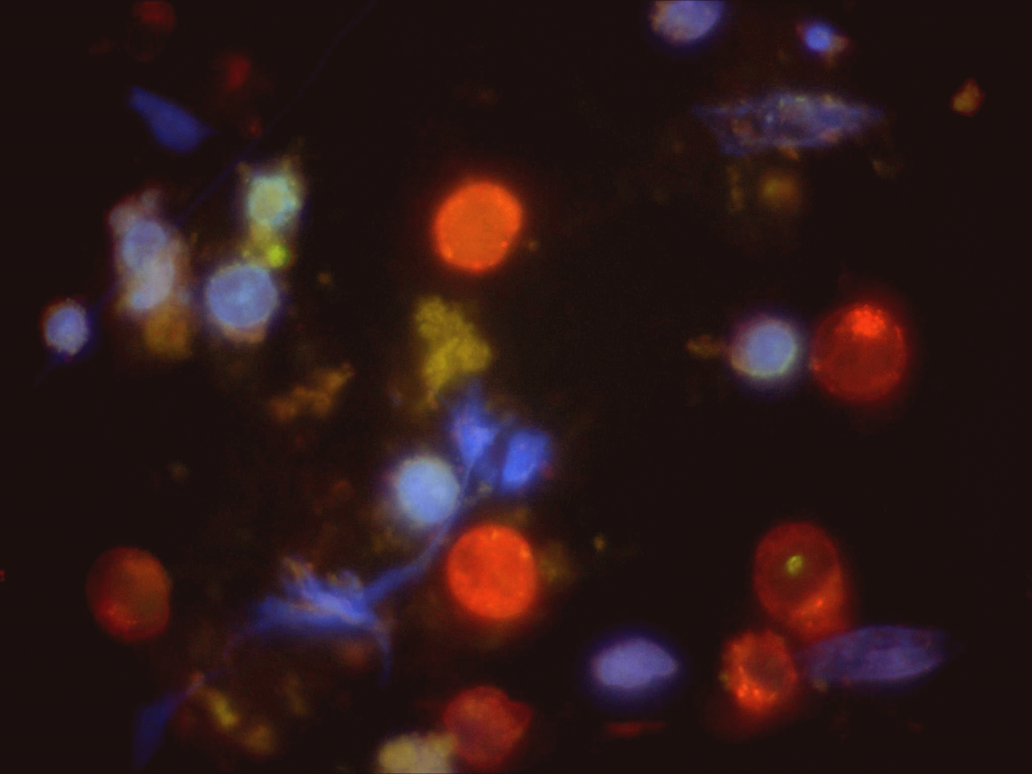
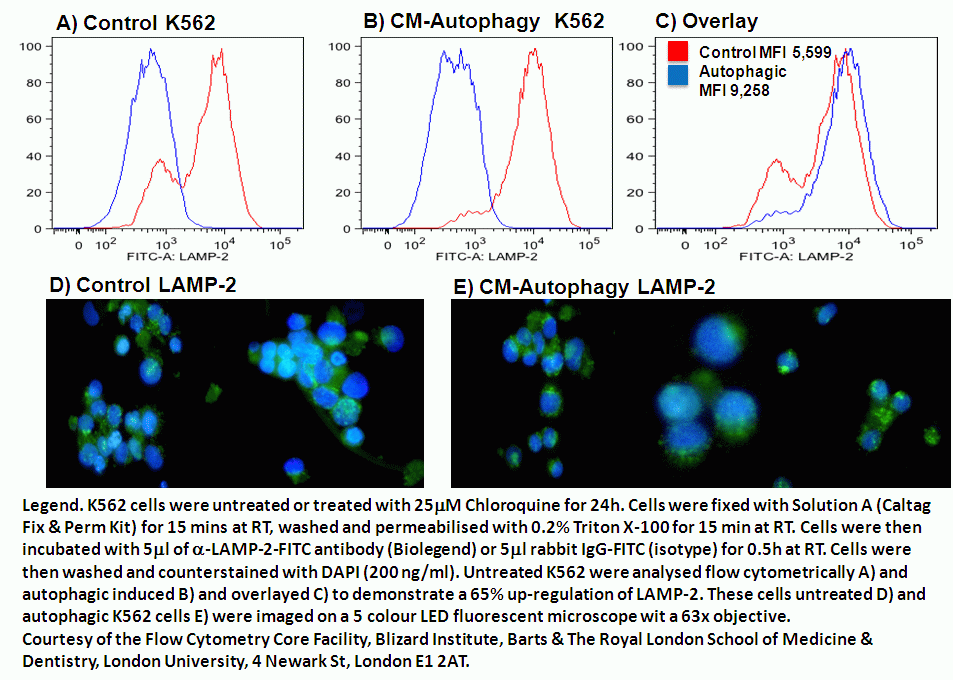
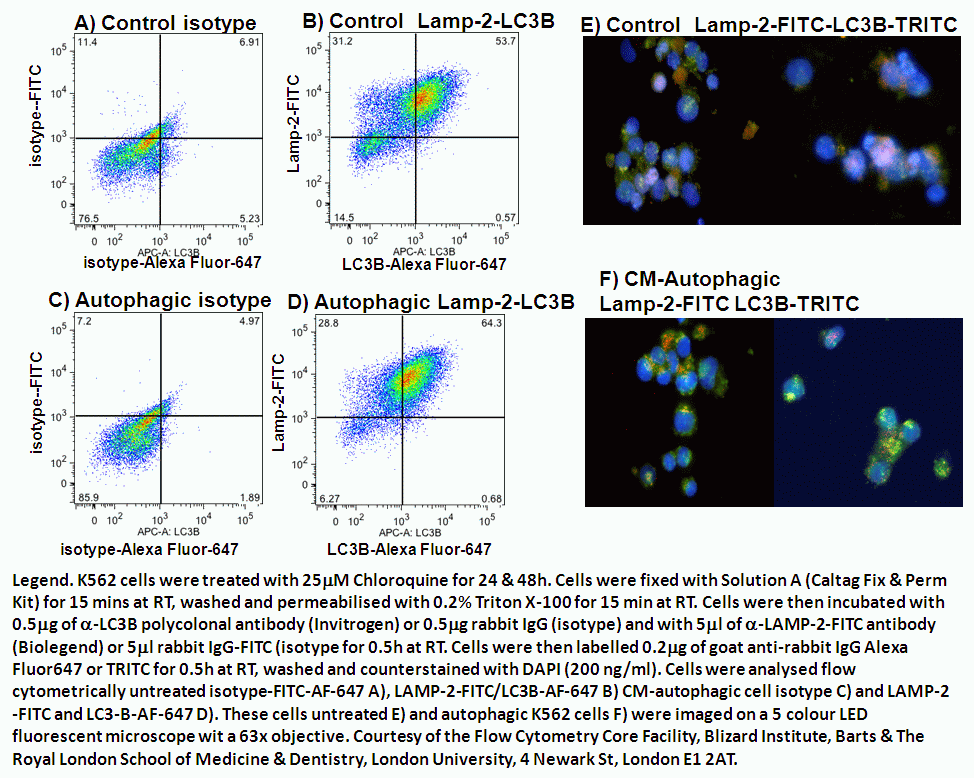
The main lysosome receptor for the clearance of cytosolic proteins via chaperone hsc70 is LAMP-2 which is up-regulated by de novo synthesis during the autophagic process, see figure. This up-regulation can be measured flow cytometrically in conjunction with the detection of the main autophagic marker, LC3B, see figure. Image analysis of such cells stained with anti-LAMP-2-FITC and anti-LC3B-TRITC clearly shows 3 populations, LC3B autophagosomes labelled red, LAMP-2 lysosomes labelled green and LC3B-LAMP-2 double positive autolysosomes labelled yellow, see figure.