
LC3B, Biological Marker for Autophagy
There are numerous methods of inducing autophagy including nutrient starvation and rapamycin which both inhibit mTOR signalling. The lack of nutrients such as serum and amino acids in the cell environment has been known to cause cell cycle arrest as well as autophagy and indeed the immunosuppressive antibiotic, rapamycin is a known to target phosphatidylinositol kinase-related kinase mTOR which combines the nutrient and mitogen signals that regulate cell cycle as well as induction of autophagy by inhibition of mTOR. Lack of serum growth factors, essential amino acids (EAA) as well as glutamine (Gln a necessary supplement for cell lines) and rapamycin has been shown to cause G1 cell cycle arrest at the restriction point (R) which is upstream of the checkpoint for EAA which is upstream of the Gln checkpoint with the rapamycin checkpoint being downstream of both of these check point just above S phase. Flow cytometry has previously been employed not only to measure LC3B but combine this with cell cycle analysis to demonstrate cell cycle arrest by rapamycin and various nutrients.
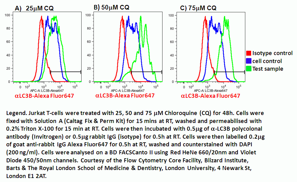
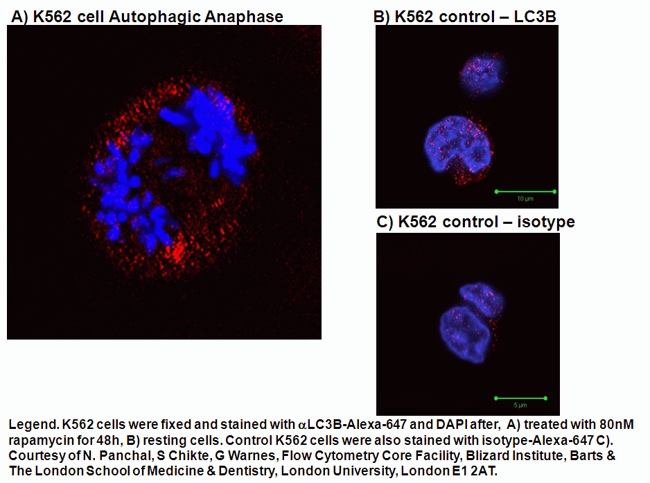
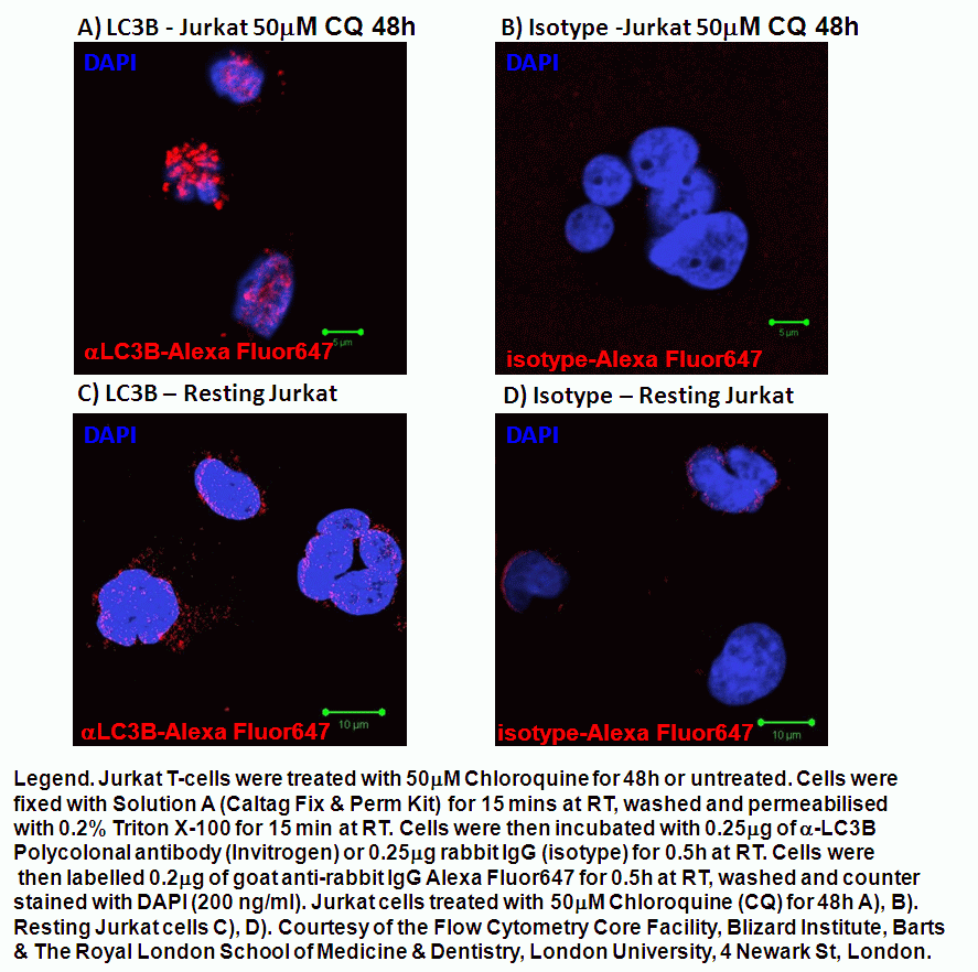
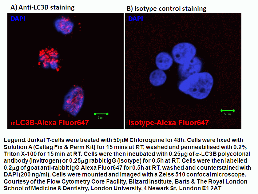
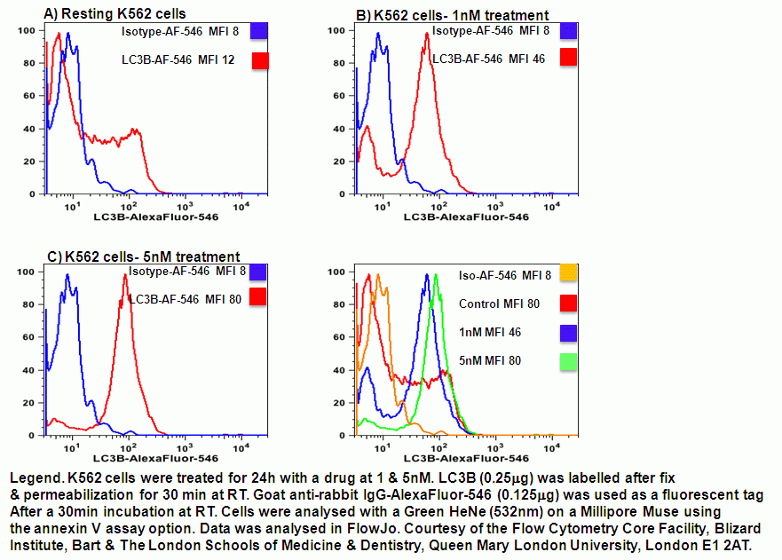
The current methods of determining the presence of autophagy is based on the fact the microtubule protein, LC3 normally present in the cytoplasm. During the autophagic process or flux LC3 is cleaved and lipidated with the phospholipid, phosphatidyl-ethanolamine (PE) to form LC3B in autophagosomes, see figure. After fusion of the autophagosome with lysosomes, to form the single membrane bound, autolysosome the the internal LC3B is reduced. The LC3B can be tagged with GFP (fluorescence is acid sensitive) and RFP (acid resistant) and imaged by fluorescence microscopy or analysed flow cytometrically. The LC3B levels can also be measured by the use of polyclonal LC3B antibody tagged in this case with Alexa Fluor 647, which can then be analysed flow cytometrically. LC3B can also be tagged with Alexa Fluor 546 and analysed using the Green HeNe laser on a Millipore Muse instrument using the Annexin V assay program, see figure.
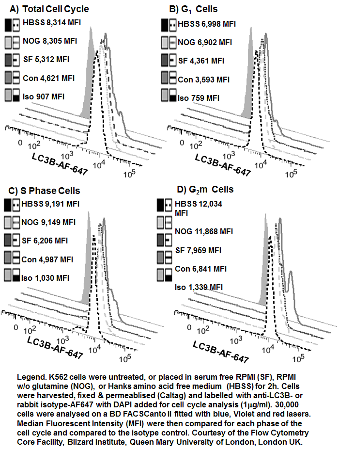
The fixation and permeabilisation of cells by the Caltag reagents allows for cell cycle analysis of autophagic cells and the level of LC3B determined for each cell cycle phase, which is shown to increase as the cell moves from G1 to S phase then to G2m. Here we show that a 2 hour serum starvation (RPMI), glutamine (RPMI w/o glutamine) and total amino acid starvation (Hanks) induces autophagy but little cell cycle arrest, see figure.
Lysosome Up-Regulation During Autophagy is Cell Cycle Dependent
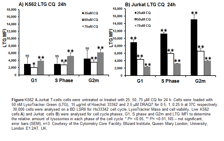
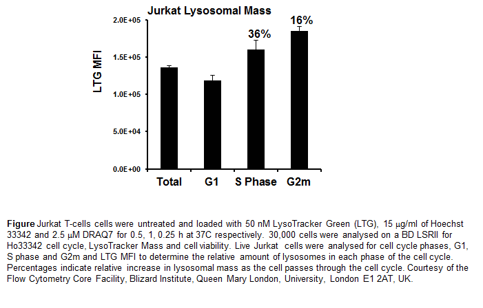
Autophagy not only up-regulates LC3B in a cell cycle dependent manner but also lysosomal mass as indicated by increased levels of LysoTracker as indicated by increasing median fluorescence intensity (MFI) in a cell cycle dependent manner, see figure. Researchers should be careful in the use of such data indicating an up-regulation of lysosomal mass during the autophagic processes as other cell mechanisms can cause an up-regulation of lysosomal mass. However in normal Jurkat T-cells lysosomal mass increases as the cell moves through the cell cycle, with S phase cells showing a 36% increase in lysosomes above that detected in cells in G1. While cells in G2m show a 16% increase in lysosomal mass above that of S phase cells, see figure.
.