
Autophagy
The process of autophagy is a cell survival mechanism that occurs when the cell is under stress, via external environmental pressures, including the lack of nutrients, or via the internal microenvironment of the cell, including the replacement of old and defective organelles such as mitochondria & the Endoplasmic Reticulum (ER).
Autophagy is also induced by the formation and collection of mis-folded proteins in the ER, which causes ER-stress within the cell. Limited autophagy of the cell allows the generation of ATP from digested organelles leading to its ultimate survival. Whilst prolonged adverse conditions results in the death of the cell by the autophagic process.
Aggresomes
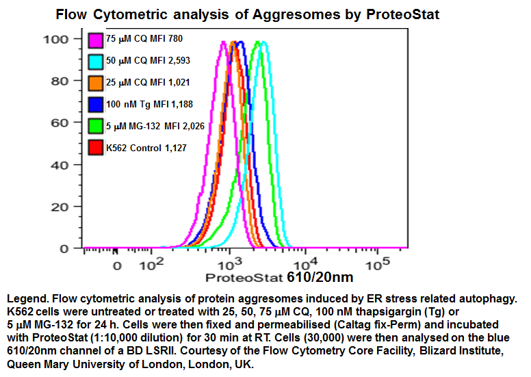
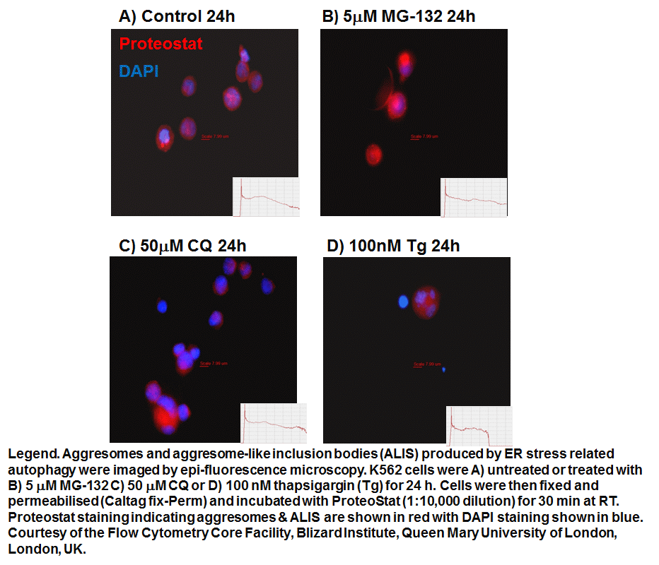
prior to macroautophagy induced ER phagy and the accumulation of mis-folded proteins within the ER, inclusion bodies form when the ubiquitin-proteasome machinery is overwhelmed by cytostolic protein aggregates to form transient structures termed Aggresome-like Induced structures or ALIS. The formation of ALIS has been shown to require signalling via mTOR and is thus independent of autophagy with ALIS being cleared by lysosomes alone. The binding of LPS or pathogens to TLR receptors not only causes NF-KB acitvation with mTOR signalling but up-regulation of p62 to form ALIS. Aggresomal responses are thought togive rise to Lewy bodies anf hyaline inclusion bodies observed in amyotrophic lateral sclerosis (ALS). Aggresosmes can be detected by use of the Proteostat Aggresome detection kit from Enzo Life Sciences which uses a red fluorescent molecular rotor dye which specifically detects denatured protein cargo within aggresomes and ALISs. Flow cytometry can used as well as fluorescent microscopy by excitation with blue laser and collection at 610nm. Protease inhibitor MG-132 can be used as a positive control for aggresome formation, see figure. Wee link to Enzo Life Sciences Website for data supplied from this Flow Cytometry Core Facility
Model of Aggresome and ALIS formation
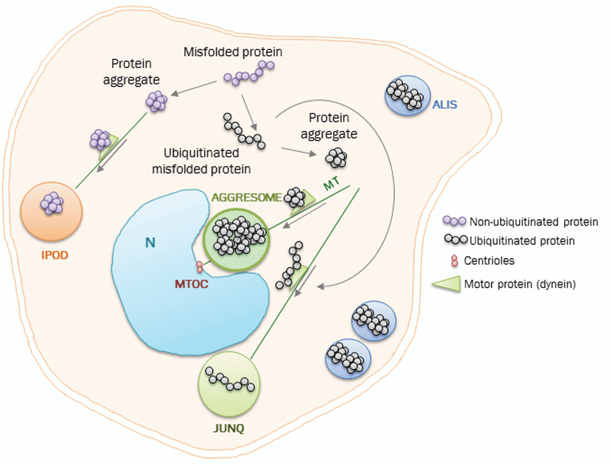
From:-Protein aggregation and degradation mechanisms in
neurodegenerative diseases by M Takalo, A Salminen, H Soininen, M Hiltunen, A Haapasalo, Am J Neurodegener Dis 2013;2(1):1-14